Research progress in the use of cationic carbon dots for the integration of cancer diagnosis with gene treatment
-
摘要: 阳离子碳点(CCDs)是表面带正电荷的一类碳点(CDs),可通过CDs和含氨基(—NH2)的阳离子化合物经“一步法”或“两步法”制备得到,既保留了CDs良好的荧光性能、低毒性及生物相容性,又提高了基因的负载输送能力及细胞摄取能力,使得CCDs在癌症靶向荧光成像和基因治疗领域中有重要的应用价值。本文综述了CCDs的制备方法和优异性能,说明CCDs可作为显像剂及基因的良好靶向载体;此外,还介绍了CCDs用于癌症诊治的基本原理及在癌症诊断和基因治疗方面的应用,并提出目前CCDs面临的问题及未来的发展趋势。Abstract: As a type of carbon dot (CD) with a positive charge on their surface, cationic carbon dots (CCDs) can be obtained from CDs and amino-containing cationic compounds by one-step or two-step preparation. They not only retain the good fluorescence performance, low toxicity and biocompatibility of CDs, but also improve their gene delivery efficiency and cell uptake capacity. These excellent properties give CCDs potential advantages in the fields of the targeted fluorescence imaging of cancers and gene therapy. This paper reviews the preparation methods and properties of CCDs, suggesting that they can be used as good targeting carriers for imaging cancer and gene therapy. In addition, the basic principles of CCDs for cancer detection and treatment, and their uses in integrated cancer diagnosis and gene therapy are introduced. Current problems and future development trends of CCDs for this purpose are discussed.
-
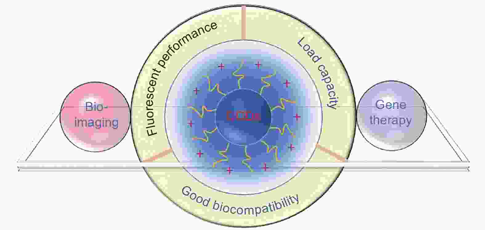
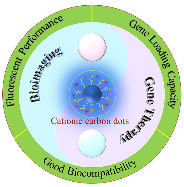
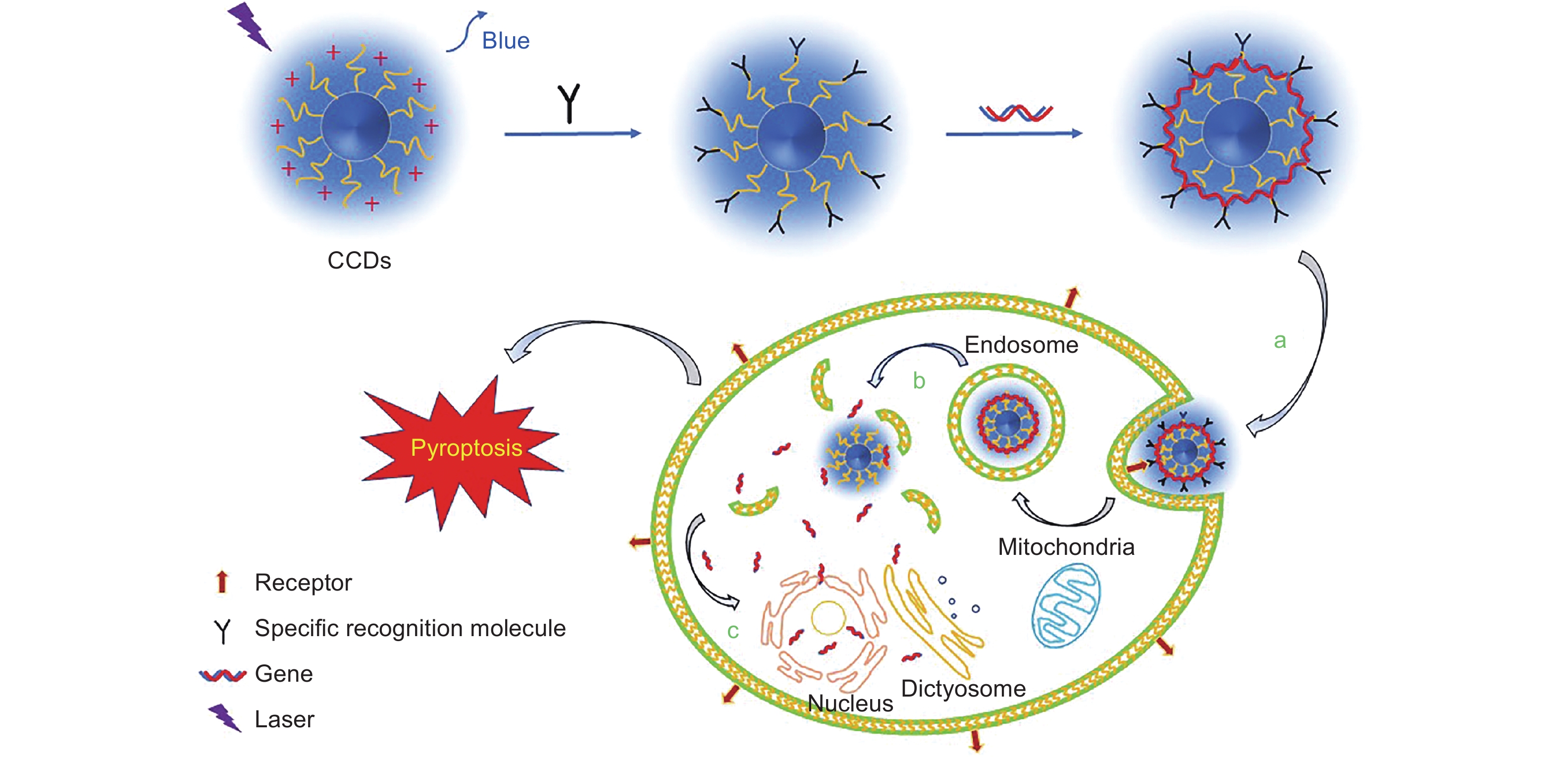
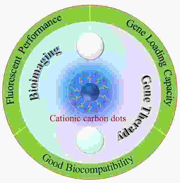
Figure 1. The properties of CCDs and their application in cancer fluorescence imaging diagnosis and gene therapy.

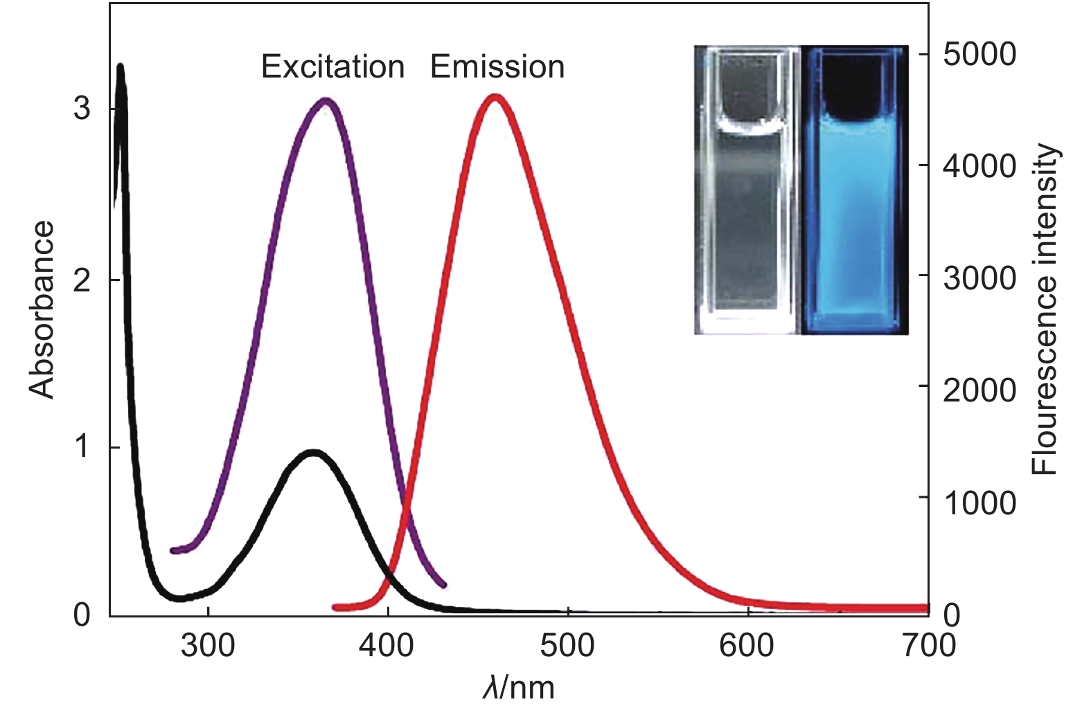
Figure 4. CCDs show good photoluminescence and fluorescence stability[47] (Reprinted with permission by copyright © 2017 American Chemical Society).
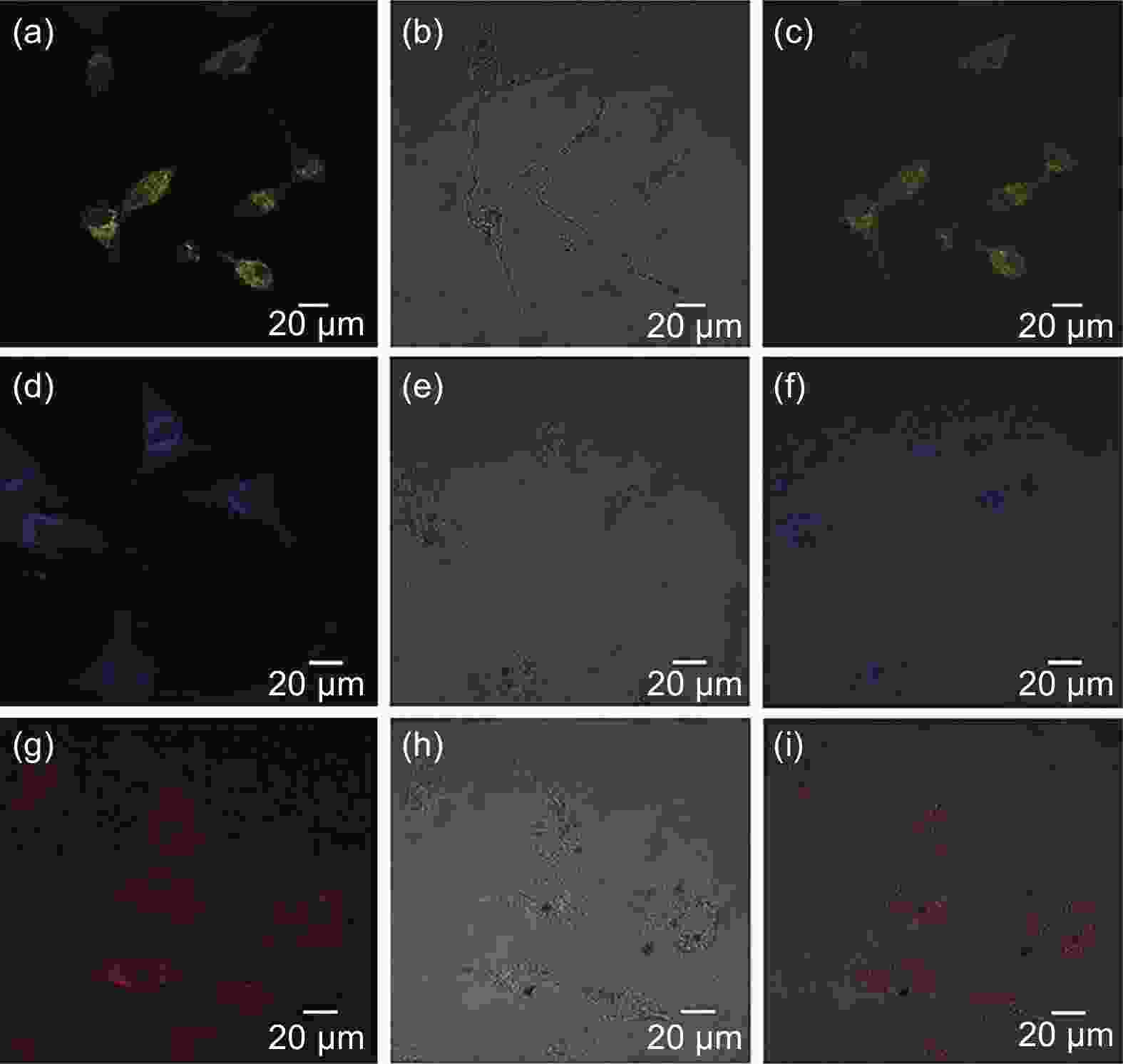
Figure 5. Confocal fluorescence images of U-87 cells incubated with (a−c) 50 μg mL−1 GQDs, (d−f) PEI1800 GQDs, and (g−i) PEI600 GQDs for 18 h. (a), (d), and (g) are the fluorescence images of U-87 cells labeled with these three GQDs, with excitation at 405 nm. (b), (e), and (h) are the bright-field images of U-87 cells. (c), (f), and (i) are the merged images of U-87 cells incubated with three GQDs[54] (Reprinted with permission by copyright © 2017 American Chemical Society).

Figure 6. (a) A) the binding capacity of CCDs and siRNA with different nitrogen/phosphate (N/P) ratios was analyzed by agarose gel electrophoresis; B) the complex formed by CCDs and siRNA with 10 nitrogen/phosphate (N/P) ratio was added to different heparin quantities for heparin decomposition analysis, (b) A−C) in vivo optical imaging, A) control group, B) free siRNA, C) CCDs/siRNA complex, and D) quantitative analysis of luciferase gene expression in fluC-4T1 xenograft after injection of CCDs/siRNA complex and (c) A−C) in vivo optical imaging, and D) quantitative analysis of luciferase gene expression in 4T1 xenograft after injection of CCDs/pDNA complex[59] (Reprinted with permission by copyright © 2014 John Wiley and Sons Ltd).

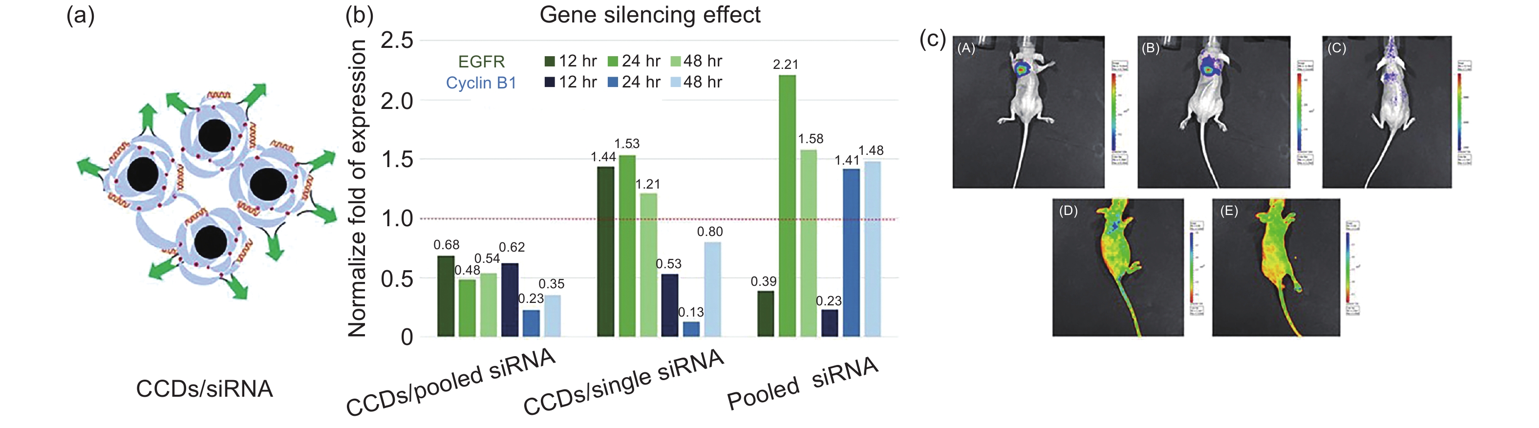
Figure 8. (a) Diagrammatic drawing of CCDs/siRNA, (b) gene silencing effect of EGFR and Cyclin B1 after CCDs/pooled siRNA, CCDs/single siRNA and pooled siRNA were treated in lung cancer H460 cells for 12, 24 and 48 h, (c) A−C) fluorescence images of lung tumors before and after treatment, B) 7 days after administration, C) 10 days after administration, D) loaded siRNA complexes deposited in the lung area, E) control group[60] (Reprinted with permission by copyright © 2016 Springer Nature).

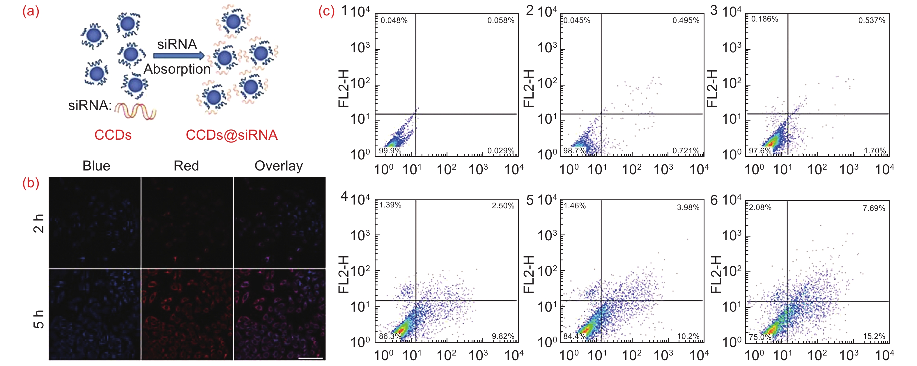
Figure 9. (a) Schematic diagram of CCDs, (b) confocal laser scanning microscope images of CCDs@siRNA and MGC-803 incubated for 2 h and 5 h, (c) apoptosis analysis of MGC-803 cells treated with CCDs@Survivin siRNA for 48 h. (1−3) are untreated group, sham transfection group and negative control group, (4−6) are Survivin siRNA-3, Survivin siRNA-2 and Survivin siRNA-1[58] (Reprinted with permission by copyright © 2014 BioMed Central).
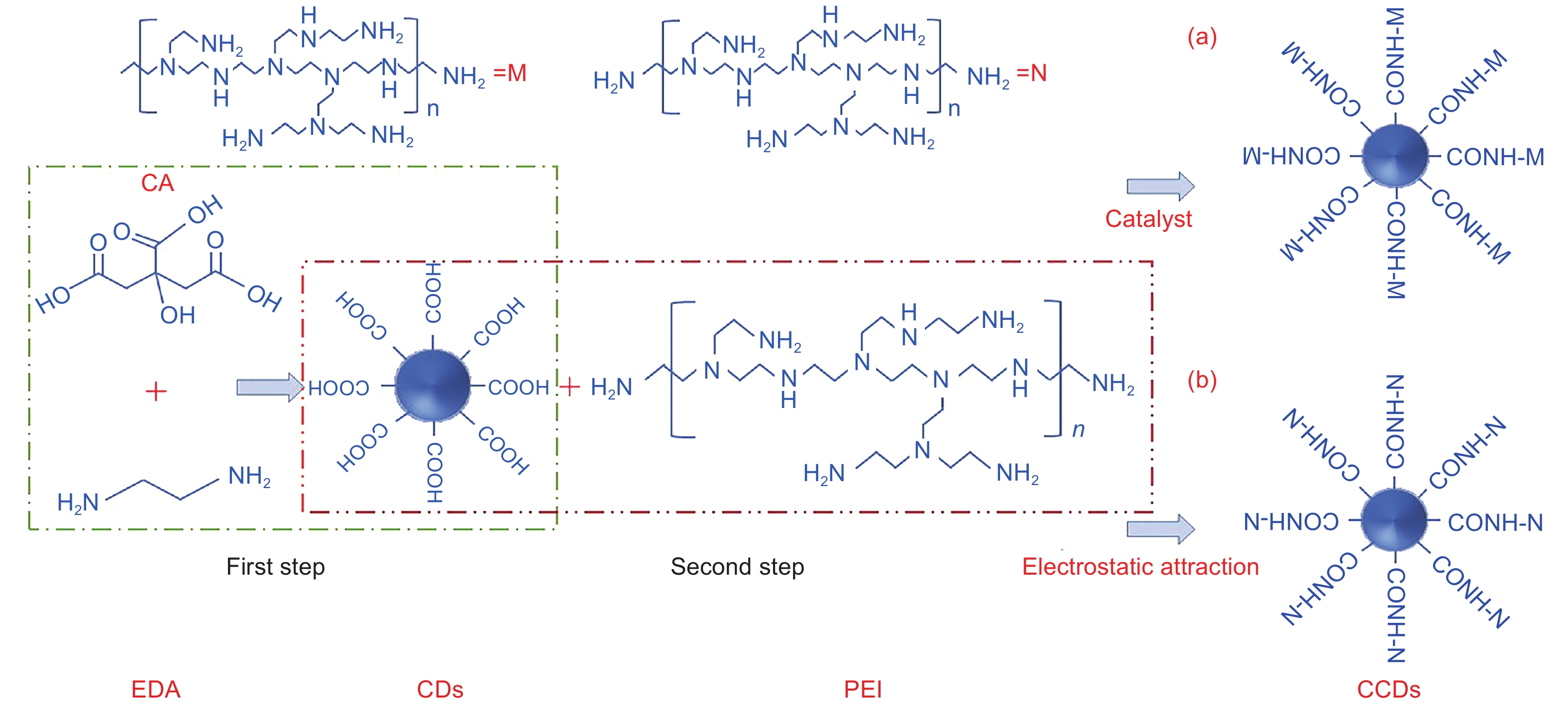
Table 1. Summary of CCDs prepared by the one-step methods.
Method Carbon source Zeta
(mV)QY
(%)Size
(nm)Fluorescent color Ref. Microwave Glucose, Arginine +21.8±0.2 12.7 1−7 Blue [32] glycerol, PEI +24 9.4 <50 Blue [33] Pyrolysis Spermidine +45 3.85 6.33±1.35 Blue [34] Hydrothermal Malic acid, PEI +26 41 1.2−5.1 Green [35] Cetrimonium bromide +10 20 1−2.5 Blue [36]  下载: 导出CSV
Table 2. Summary of CCDs prepared by the two-step methods.
Method Carbon source Zeta (mV) QY (%) Size (nm) Fluorescent color Ref. Amide reaction CA, Urea, bPEI +24 18.7 10 Blue [37] CA, EDA, bPEI +46.68±
0.78- 22.8±6.7 Blue [38] Electrostatic attraction Candle soot, PEI +40 - <8.1 - [39] CA, EDA, PEI +15.6 - 122.7 Blue [40]
下载: 导出CSV
-
[1] WANG Ning, LIU Shuo, YANG Lei, et al. Interpretation of the 2018 global cancer statistics report[J]. Journal of Multidisciplinary Cancer Management (Electronic Version),2019,5(01):87-97. [2] Ren W, Yu J, Zhang Z M, et al. Missed diagnosis of early gastric cancer or high-grade intraepithelial neoplasia[J]. World Journal of Gastroenterology,2013(13):2092-2096. [3] Rembacken B J, Fujii T, Cairns A, et al. Flat and depressed colonic neoplasms: a prospective study of 1000 colonoscopies in the UK[J]. Lancet,2000,355:1211-1214. doi: 10.1016/S0140-6736(00)02086-9 [4] NIE De-hong, LI Hong-yi, LI Kai, et al. The present situation and new progress of imaging examination of breast cancer[J]. China Modern Medicine,2015,22(15):20-24. [5] Ferlay J, Soerjomataram I, Dikshit R, et al. Cancer incidence and mortality worldwide: sources, methods and major patterns in GLOBOCAN 2012[J]. International Journal of Cancer,2015,136(5):E359-E386. doi: 10.1002/ijc.29210 [6] Mohanti B K, Rath G K, Anantha N, et al. Improving cancer radiotherapy with 2-deoxy-D-glucose: phase I/Ⅱ clinical trials on human cerebral gliomas[J]. International Journal of Radiation Oncology, Biology, Physics,1996,35(1):103-111. doi: 10.1016/S0360-3016(96)85017-6 [7] Yang J Y, Ju Z J, Dong S F. Cisplatin and paclitaxel co-delivered by folate-decorated lipid carriers for the treatment of head and neck cancer[J]. Drug Delivery,2016,24(1):792-799. [8] Ishiwata H, Suzuki N, Ando S, et al. Characteristics and biodistribution of cationic liposomes and their DNA complexes[J]. Journal of Controlled Release,2000,69(1):139-148. doi: 10.1016/S0168-3659(00)00293-5 [9] Niven R, Pearlman R, Wedeking T, et al. Biodistribution of radiolabeled lipid-DNA complexes and DNA in mice[J]. Journal of pharmaceutical sciences,1998,87:1292-1299. doi: 10.1021/js980087a [10] Hu Z, Zhang D Y, Lu S T, et al. Chitosan-based composite materials for prospective hemostatic applications[J]. Marine Drugs,2018,16(8):273. doi: 10.3390/md16080273 [11] Misra S K, Naz S, Kondaiah P, et al. A cationic cholesterol based nanocarrier for the delivery of p53-EGFP-C3 plasmid to cancer cells[J]. Biomaterials,2014,35(4):1334-1346. doi: 10.1016/j.biomaterials.2013.10.062 [12] Hua X W, Bao Y W, Chen Z, et al. Carbon quantum dots with intrinsic mitochondrial targeting ability for mitochondria-based theranostics[J]. Nanoscale,2017,9(30):10948-10960. doi: 10.1039/C7NR03658B [13] WEI Ying-ying, CHEN Lin, WANG Jun-li, et al. Synthesis and applications of chiral carbon quantum dots[J]. Progress in Chemistry,2020,32(04):381-391. [14] Lu C X, Li L P. Progress in research on the preparation of carbon dots and their use in tumor theranostics[J]. New Carbon Materials,2018,33(1):12-18. [15] Liu W, Zhang R, Kang Y, et al. Preparation of nitrogen-doped carbon dots with a high fluorescence quantum yield for the highly sensitive detection of Cu2+ ions, drawing anti-counterfeit patterns and imaging live cells[J]. New Carbon Materials,2019,34(04):390-402. doi: 10.1016/S1872-5805(19)30024-1 [16] Yu S P, Su X D, Du J L, et al. The cytotoxicity of water-soluble carbon nanotubes on human embryonic kidney and liver cancer cells[J]. New Carbon Materials,2018,33(1):36-45. doi: 10.1016/S1872-5805(18)60325-7 [17] Zheng J X, Liu X H, Yang Y Z, et al. Rapid and green synthesis of fluorescent carbon dots from starch for white light-emitting diodes[J]. New Carbon Materials,2018,33(03):276-288. doi: 10.1016/S1872-5805(18)60339-7 [18] Kayo O V, Jefferson B, Luiz F C D O, et al. Synthesis of multicolor photoluminescent carbon quantum dots functionalized with hydrocarbons of different chain lengths[J]. New Carbon Materials,2017,32(04):327-337. doi: 10.1016/S1872-5805(17)60126-4 [19] Cui B, Feng X T, Zhang F, et al. The use of carbon quantum dots as fluorescent materials in white LEDs[J]. New Carbon Materials,2017,32(05):385-401. doi: 10.1016/S1872-5805(17)60130-6 [20] Liu Y, Li Q Q, Zhang H, et al. Research progress on the use of micro/nano carbon materials for antibacterial dressings[J]. New Carbon Materials,2020,35(04):323-335. doi: 10.1016/S1872-5805(20)60492-9 [21] Mao X J, Zheng H Z, Long Y J, et al. Study on the fluorescence characteristics of carbon dots[J]. Spectrochimica Acta Part A: Molecular and Biomolecular Spectroscopy,2010,75(2):553-557. doi: 10.1016/j.saa.2009.11.015 [22] Xu X Y, Ray R, Gu Y L, et al. Electrophoretic analysis and purification of fluorescent single-walled carbon nanotube fragments[J]. Journal of the American Chemical Society,2004,126(40):12736-12737. doi: 10.1021/ja040082h [23] Wang X, Cao L, Lu F S, et al. Photoinduced electron transfers with carbon dots[J]. Chemical Communications,2009,46(25):3774-3776. [24] Zheng L Y, Chi Y W, Dong Y Q, et al. Electrochemiluminescence of water-soluble carbon nanocrystals released electrochemically from graphite[J]. Journal of the American Chemical Society,2009,131(13):4564-4565. doi: 10.1021/ja809073f [25] Cao L, Wang X, Meziani M J, et al. Carbon dots for multiphoton bioimaging[J]. Journal of the American Chemical Society,2007,129(37):11318-11319. doi: 10.1021/ja073527l [26] Tripathi K M, Sonker A K, Sonkar S K, et al. Pollutant soot of diesel engine exhaust transformed to carbon dots for multicoloured imaging of E. coli and sensing cholesterol[J]. RSC Advances,2014,4(57):30100-30107. doi: 10.1039/C4RA03720K [27] Hu G K, Ge L, Li Y Y, et al. Carbon dots derived from flax straw for highly sensitive and selective detections of cobalt, chromium, and ascorbic acid[J]. Journal of Colloid and Interface Science,2020,579:96-108. doi: 10.1016/j.jcis.2020.06.034 [28] Zhu H, Wang X L, Li Y L, et al. Microwave synthesis of fluorescent carbon nanoparticles with electrochemiluminescence properties[J]. Chemical Communications,2009,1(34):5118-5120. [29] Bourlinos A B, Stassinopoulos A, Anglos D, et al. Surface functionalized carbogenic quantum dots[J]. Small,2008,4(4):455-458. doi: 10.1002/smll.200700578 [30] Liu R L, Wu D Q, Liu S H, et al. An aqueous route to multicolor photoluminescent carbon dots using silica spheres as carriers[J]. Angewandte Chemie International Edition in English,2009,48(25):4598-4601. doi: 10.1002/anie.200900652 [31] Hou P, Yang T, Liu H, et al. An active structure preservation method for developing functional graphitic carbon dots as an effective antibacterial agent and a sensitive pH and Al(Ⅲ) nanosensor[J]. Nanoscale,2017,9(44):17334-17341. doi: 10.1039/C7NR05539K [32] Cao X, Wang J P, Deng W W, et al. Photoluminescent cationic carbon dots as efficient non-viral delivery of plasmid SOX9 and chondrogenesis of fibroblasts[J]. Scientific Reports,2018,8(1):1-11. doi: 10.1038/s41598-017-17765-5 [33] Liu C J, Zhang P, Zhai X Y, et al. Nano-carrier for gene delivery and bioimaging based on carbon dots with PEI-passivation enhanced fluorescence[J]. Biomaterials,2012,33(13):3604-3613. doi: 10.1016/j.biomaterials.2012.01.052 [34] Jian H J, Wu R S, Lin T Y, et al. Super-cationic carbon quantum dots synthesized from spermidine as an eye drop formulation for topical treatment of bacterial keratitis[J]. ACS Nano,2017,11(7):6703-6716. doi: 10.1021/acsnano.7b01023 [35] Guo Y, Zhang J C, Zhang W H, et al. Green fluorescent carbon quantum dots functionalized with polyethyleneimine, and their application to aptamer-based determination of thrombin and ATP[J]. Microchimica Acta,2019,186(11):1-8. [36] Saberi Z, Rezaei B, Ensafi A A. Fluorometric label-free aptasensor for detection of the pesticide acetamiprid by using cationic carbon dots prepared with cetrimonium bromide[J]. Microchimica Acta,2019,186(5):1-7. [37] Cui H Y. Integrated carbon quantum dots for cancer cell targeted fluorescence imaging and gene therapy[D]. Dalian University of Technology, 2018. [38] Shu M J, Gao F, Yu C L, et al. Dual targeted therapy in HER2-positive breast cancer cells with the combination of carbon dots/HER3 siRNA and trastuzumab[J]. Nanotechnology,2020,31(33):335102. doi: 10.1088/1361-6528/ab8a8a [39] Liu Y, Hu J, Li Y, et al. Synthesis of polyethyleneimine capped carbon dots for preconcentration and slurry sampling analysis of trace chromium in environmental water samples[J]. Talanta,2015,134:16-23. doi: 10.1016/j.talanta.2014.11.001 [40] Kong T, Zhou R, Zhang Y, et al. AS1411 aptamer modified carbon dots via polyethylenimine-assisted strategy for efficient targeted cancer cell imaging[J]. Cell Proliferation,2020,53(1):e12713. [41] Periasamy A. Optical imaging techniques in cell biology[J]. Microscopy and Microanalysis,2013,19(4):1106. doi: 10.1017/S1431927613001645 [42] Abdallah B, Hassan A, Benoist C, et al. A powerful nonviral vector for in vivo gene transfer into the adult mammalian brain: Polyethylenimine[J]. Human Gene Therapy,1996,7(16):1947-1954. doi: 10.1089/hum.1996.7.16-1947 [43] Hasanzadeh A, Radmanesh F, Kiani J, et al. Photoluminescent functionalized carbon dots for CRISPR delivery: synthesis, optimization and cellular investigation[J]. Nanotechnology,2019,30(13):135101. doi: 10.1088/1361-6528/aafbf9 [44] Dong Y Q, Wang R X, Li H, et al. Polyamine-functionalized carbon quantum dots for chemical sensing[J]. Carbon,2012,50(8):2810-2815. doi: 10.1016/j.carbon.2012.02.046 [45] Nirmal M, Dabbousi B, Bawendi M G, et al. Fluorescence intermittency in single cadmium selenide nanocrystals[J]. Nature,1996,383(6603):802-804. doi: 10.1038/383802a0 [46] Yang X D, Wang Y, Shen X R, et al. One-step synthesis of photoluminescent carbon dots with excitation-independent emission for selective bioimaging and gene delivery[J]. Journal of Colloid and Interface Science,2017,492:1-7. doi: 10.1016/j.jcis.2016.12.057 [47] Li J Y, Liu Y, Shu Q W, et al. One-pot hydrothermal synthesis of carbon dots with efficient up- and down-converted photoluminescence for the sensitive detection of morin in a dual-readout assay[J]. Langmuir,2017,33(4):1043-1050. doi: 10.1021/acs.langmuir.6b04225 [48] Wu L L, Li X L, Ling Y F, et al. Morpholine derivative-functionalized carbon dots-based fluorescent probe for highly selective lysosomal imaging in living cells[J]. ACS Applied Materials & Interfaces,2017,9(34):28222-28232. [49] Yuan Y H, Liu Z X, Li R S, et al. Synthesis of nitrogen-doping carbon dots with different photoluminescence properties by controlling the surface states[J]. Nanoscale,2016,8(12):6770-6776. doi: 10.1039/C6NR00402D [50] Sun Y P, Zhou B, Lin Y, et al. Quantum-sized carbon dots for bright and colorful photoluminescence[J]. Journal of the American Chemical Society,2006,128(24):7756-7757. doi: 10.1021/ja062677d [51] Han B F, Wang W X, Wu H Y, et al. Polyethyleneimine modified fluorescent carbon dots and their application in cell labeling[J]. Colloids and Surfaces B: Biointerfaces,2012,100:209-214. doi: 10.1016/j.colsurfb.2012.05.016 [52] Arsalani N, Nezhad-Mokhtari P, Jabbari E. Microwave-assisted and one-step synthesis of PEG passivated fluorescent carbon dots from gelatin as an efficient nanocarrier for methotrexate delivery[J]. Artificial Cells Nanomedicine and Biotechnology,2019,47(1):540-547. doi: 10.1080/21691401.2018.1562460 [53] Liu H, Ye T, Mao C. Fluorescent carbon nanoparticles derived from candle soot[J]. Angewandte Chemie,2007,46(34):6593-6595. [54] Gao T, Wang X, Yang L Y, et al. Red, yellow and blue luminescence by graphene quantum dots: Syntheses, mechanism, and cellular imaging[J]. ACS Applied Materials & Interfaces,2017,9(29):24846-24856. [55] Shahid S, Mohiyuddin S, Packirisamy G. Synthesis of multi-color fluorescent carbon dots from mint leaves: A robust bioimaging agent with potential antioxidant activity[J]. Journal of Nanoscience and Nanotechnology,2020,20(10):6305-6316. doi: 10.1166/jnn.2020.17899 [56] WANG Jun-li, WANG Ya-ling, ZHENG Jing-xia, et al. Mechanism, tuning and application of excitation-dependent fluorescence property in carbon dots[J]. Progress in Chemistry,2018,30(08):1186-1201. [57] Neu M, Fischer D, Kissel T. Recent advances in rational gene transfer vector design based on poly(ethylene imine) and its derivatives[J]. Journal of Gene Medicine,2005,7(8):992-1009. doi: 10.1002/jgm.773 [58] Wang Q, Zhang C L, Shen G X, et al. Fluorescent carbon dots as an efficient siRNA nanocarrier for its interference therapy in gastric cancer cells[J]. Journal of Nanobiotechnology,2014,12(1):58. doi: 10.1186/s12951-014-0058-0 [59] Wang L Q, Wang X Y, Bhirde A, et al. Carbon-dot-based two-photon visible nanocarriers for safe and highly efficient delivery of siRNA and DNA[J]. Advanced Healthcare Materials,2014,3(8):1203-1209. doi: 10.1002/adhm.201300611 [60] Wu Y F, Wu H C, Kuan C H, et al. Multi-functionalized carbon dots as theranostic nanoagent for gene delivery in lung cancer therapy[J]. Scientific Reports,2016,6:21170. doi: 10.1038/srep21170 [61] Wang H J, He X, Luo T Y, et al. Amphiphilic carbon dots as versatile vectors for nucleic acid and drug delivery[J]. Nanoscale,2017,9(18):5935-5947. doi: 10.1039/C7NR01029J [62] Dong H, Shim K N, Li J M J, et al. Molecular mechanisms underlying Ca2+ mediated motility of human pancreatic duct cells[J]. American Journal of Physiology Cell Physiology,2010,299(6):C1493-1503. doi: 10.1152/ajpcell.00242.2010 [63] Matejcic M, De B J, Ricci C, et al. Biomarkers of folate and vitamin B12 and breast cancer risk: Report from the EPIC cohort[J]. International Journal of Cancer,2017,140(6):1246-1259. doi: 10.1002/ijc.30536 [64] Sahay G, Alakhova D Y, Kabanov A V. Endocytosis of nanomedicines[J]. Journal of Controlled Release,2010,145(3):182-195. doi: 10.1016/j.jconrel.2010.01.036 [65] McMahon H T, Boucrot E. Molecular mechanism and physiological functions of clathrin-mediated endocytosis[J]. Nature Reviews Molecular Cell Biology,2011,12:517-533. doi: 10.1038/nrm3151 [66] Gratton S E, Ropp P A, Pohlhaus P D, et al. The effect of particle design on cellular internalization pathways[J]. Proceedings of the National Academy of Sciences of the United States of America,2008,105(33):11613-11618. doi: 10.1073/pnas.0801763105 [67] Zhou J, Deng W, Wang Y, et al. Cationic carbon quantum dots derived from alginate for gene delivery: One-step synthesis and cellular uptake[J]. Acta Biomaterialia,2016,42:209-219. doi: 10.1016/j.actbio.2016.06.021 [68] Averykiejda K A, Bowden N A, Croft A, et al. P53 in human melanoma fails to regulate target genes associated with apoptosis and the cell cycle and may contribute to proliferation[J]. BMC Cancer,2011,11(1):203-203. doi: 10.1186/1471-2407-11-203 [69] Svoboda P, Stein P, Hayashi H, et al. Selective reduction of dormant maternal mRNAs in mouse oocytes by RNA interference[J]. Development,2000,127(19):4147-4156. [70] Wu D, Li B L, Zhao Q W, et al. Assembling defined DNA nanostructure with nitrogen-enriched carbon dots for theranostic cancer applications[J]. Small,2020,16(19):e1906975. doi: 10.1002/smll.201906975 [71] Du F, Zhao X, Lu W, et al. Dual-ligand functionalized carbon nanodots as green fluorescent nanosensors for cellular dual receptor-mediated targeted imaging[J]. Analyst,2019,144(22):6729-6735. doi: 10.1039/C9AN01530B [72] Behr J P. The proton sponge: A trick to enter cells the viruses did not exploit[J]. Chimia International Journal for Chemistry,1997,51(1/2):34-36. -


 点击查看大图
计量
- 文章访问数: 907
- HTML全文浏览量: 613
- PDF下载量: 112
- 被引次数: 0