Atomic-scale investigation of carbon-based materials by gentle transmission electron microscopy
-
摘要: 近年来,石墨烯、金属有机框架(MOFs)、聚合物、生物大分子等碳基材料在物理、化学、材料、生物学等领域受到了持续增长的关注,但由于其微观结构在电子束辐照下的不稳定性导致原子分辨率的实验观察仍面临巨大的挑战。原子结构的认识不足严重限制了对此类材料的深入理解及潜在应用研究。近年来,低加速电压、低电子剂量与低温电子显微学(TEM)亚埃分辨率的革命性突破,极大地促进了对电子束敏感材料结构与化学组成的原子尺度解析。特别是对轻元素原子成像的能力有助于深入研究新型碳基材料的结构和性能。本文总结了先进电子显微学中各种成像与谱学技术的新进展,及其在石墨烯基材料、MOFs、聚合物、生物大分子等碳基材料研究中的应用,并探讨了当前材料研究面临的挑战与电子显微学的发展趋势,以期助力对新型碳基材料构效关系的深入理解和设计开发。Abstract: Although carbon-based materials, such as graphene, metal-organic frameworks (MOFs), polymers and biomolecules, have aroused increasing scientific interest in the fields of physics, chemistry, materials science and molecular biology, their atomic-scale observation is still a challenge due to their structural instability under the electron beam. Ambiguous atomic arrangements have critically limited the fundamental understanding on these materials and their potential applications in electronics, mechanics, thermodynamics, catalysis, bioscience and medicine. Very recently, revolutionary sub-Ångström resolution achievements of transmission electron microscopy (TEM) using a low voltage, a low electron dose, or a cryogenic environment have greatly facilitated the atomic-scale structural and chemical examination of electron beam sensitive materials. In particular, the ability to image light elements atom by atom gives unprecedented insight into the structures and properties of novel carbon-based materials. In this review, the recent developments in advanced TEM combined with various imaging and spectroscopy techniques, and their use in examining graphene-based materials, MOFs, polymers, and biomacromolecules are summarized and discussed. The current challenges in materials research and trends for the future design of TEM equipment are outlined, which is expected to provide a deeper understanding of structure–performance relationships and the discovery of new carbon materials.
-
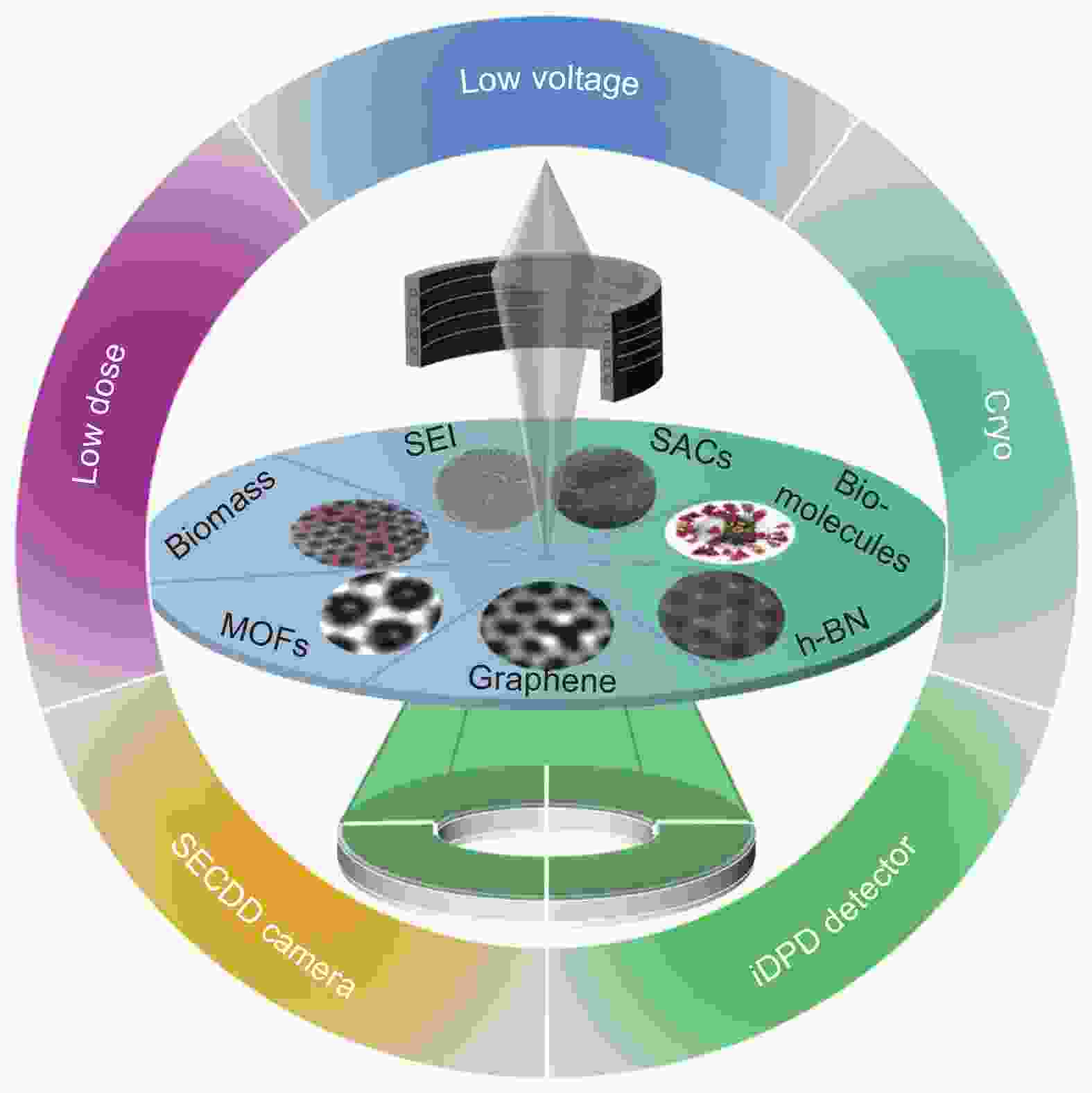
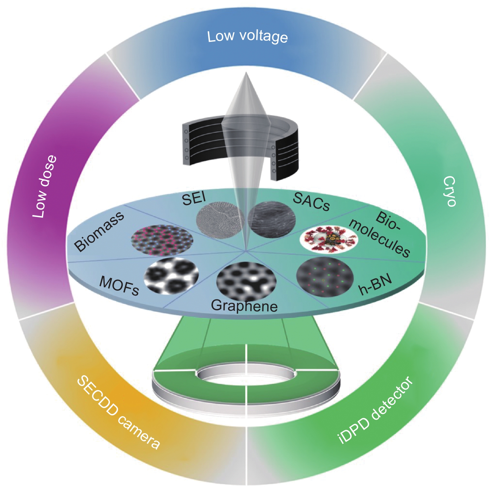
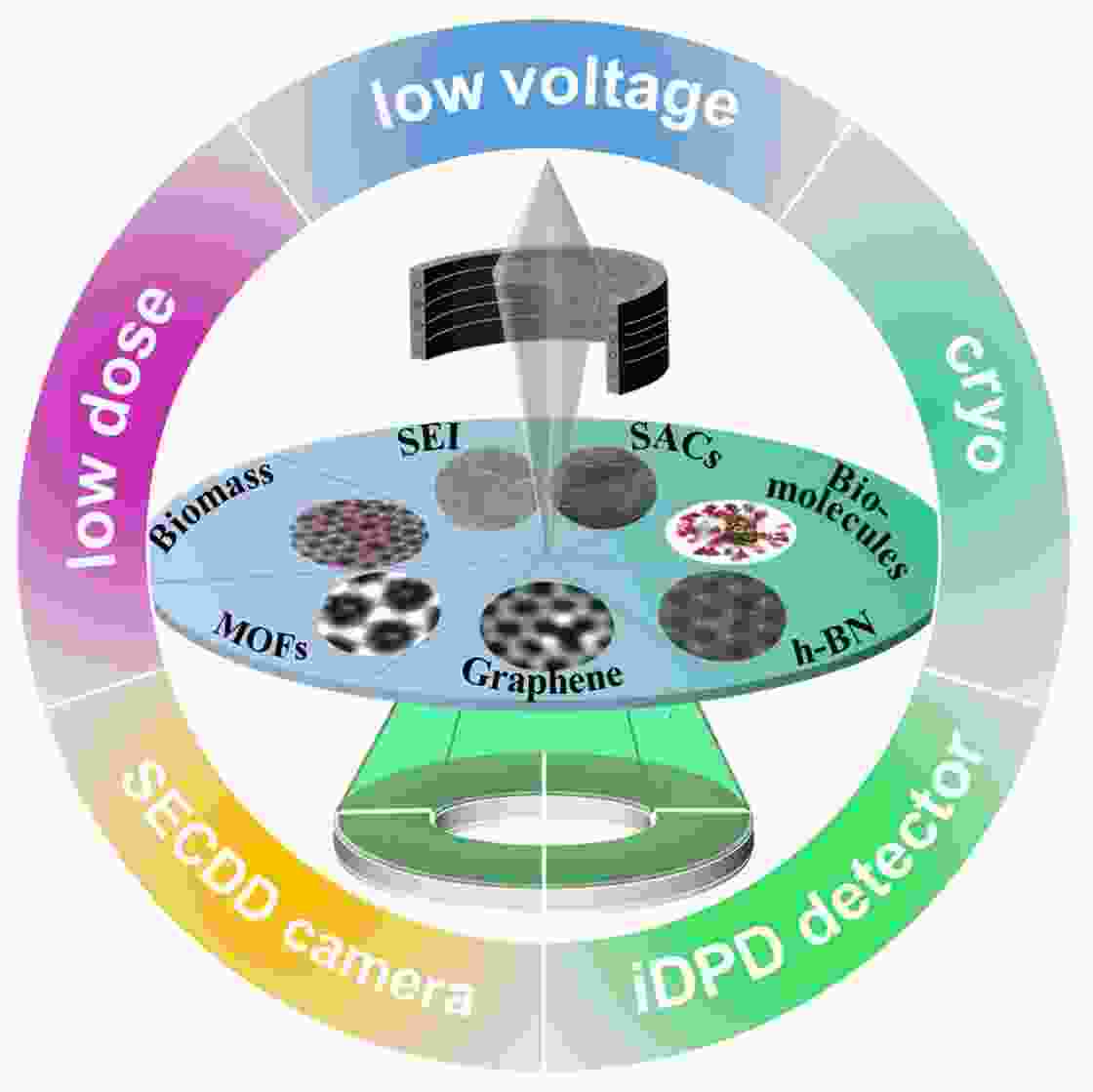
Figure 1. A summary of Cs corrected TEM techniques applied in novel carbon-based materials.
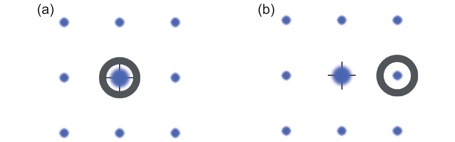
Figure 3. Diagrams illustrated (a) BF and (b) DF illumination arrangements in a conventional TEM. Faint spots denote diffraction dots, the crosshair in the center denotes the TEM optic axis, and the dark rings denote the objective aperture.
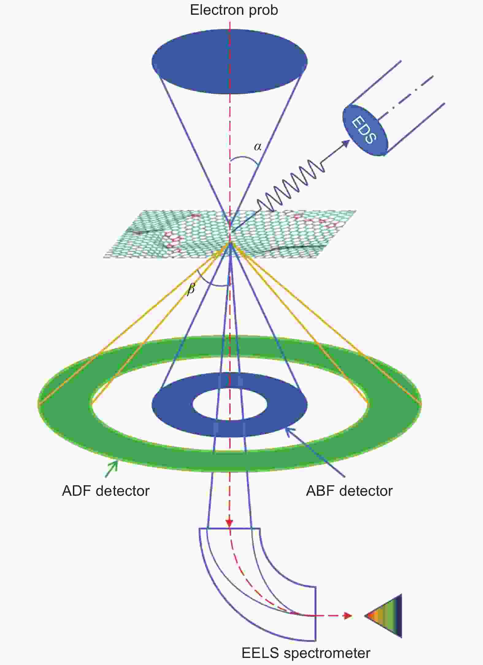
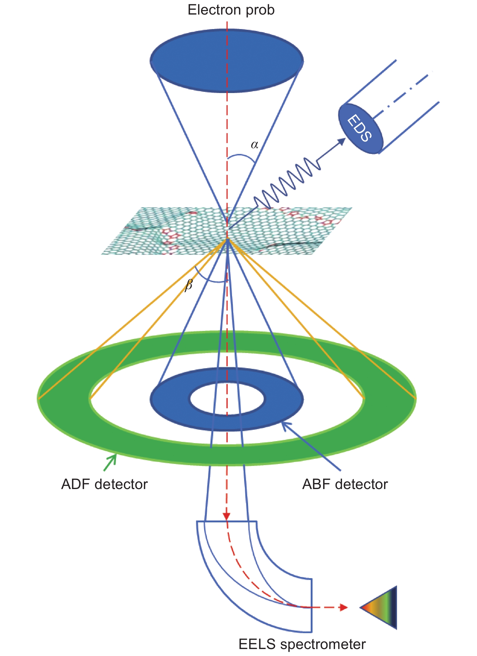
Figure 4. Schematic diagram of the common imaging and spectrum modes in a STEM, composed of ADF or ABF imaging, EDX, and EELS.
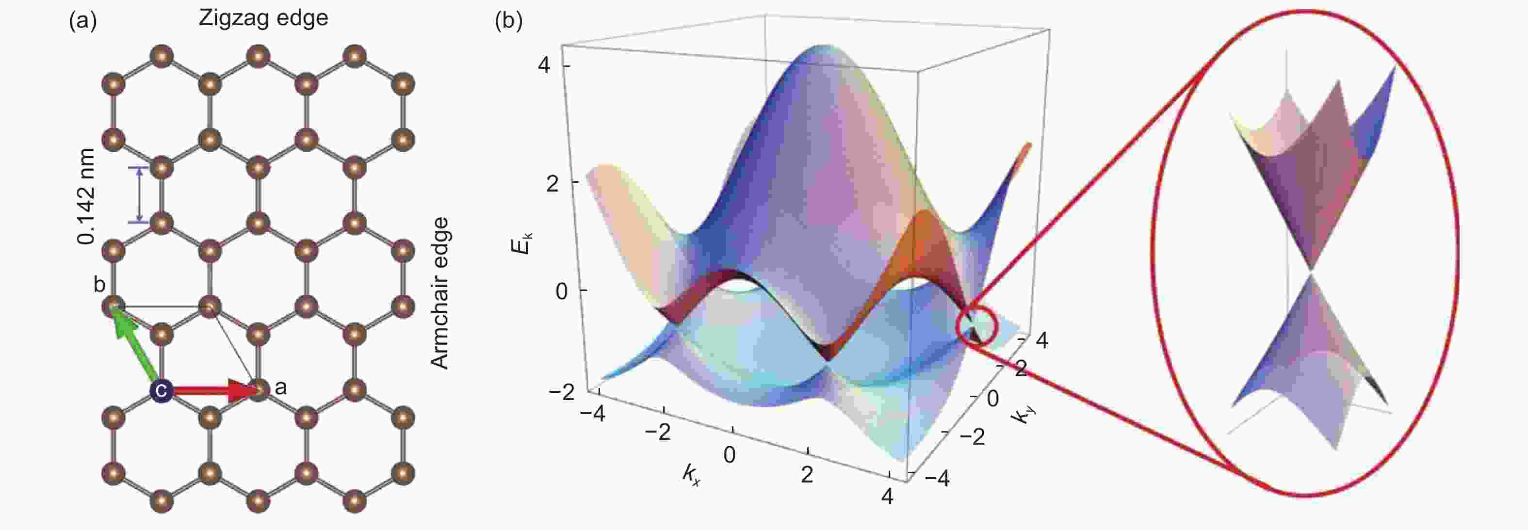
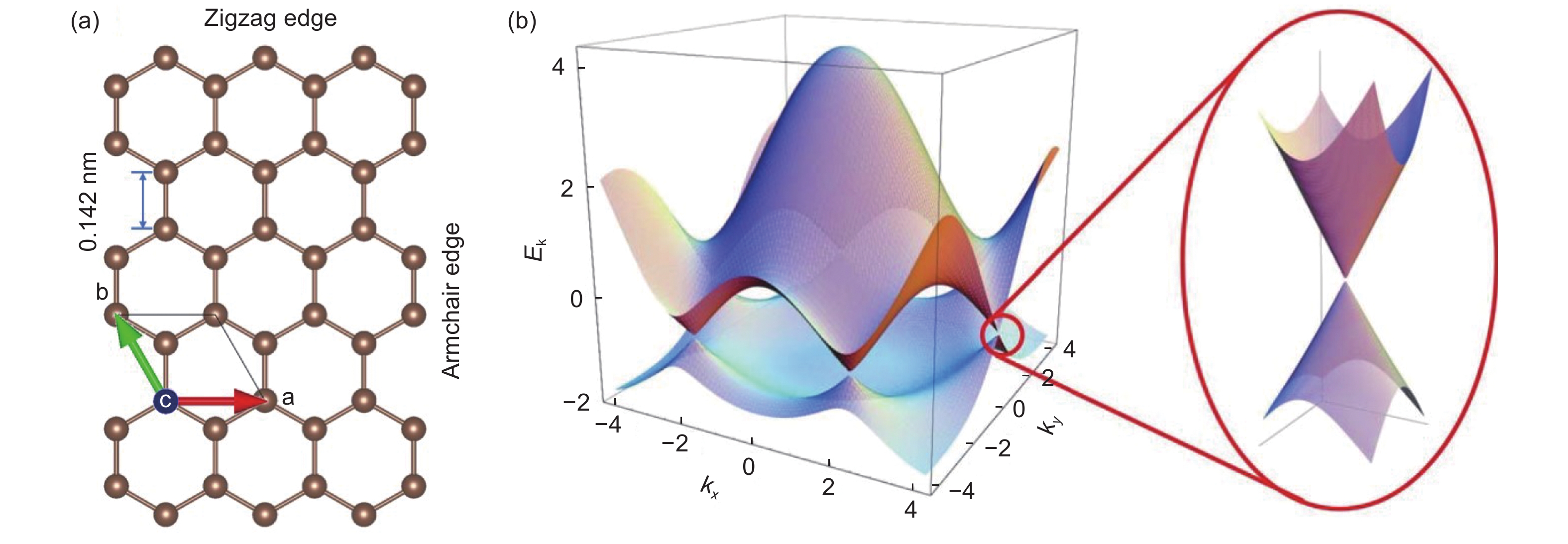
Figure 5. The structure of graphene: (a) Honeycomb structure of graphene, with a hexagonal unit cell and a bond length of 0.142 nm. Graphene has 2 types of common edges: the zigzag edge and armchair edge, (b) Band structure of graphene[46].

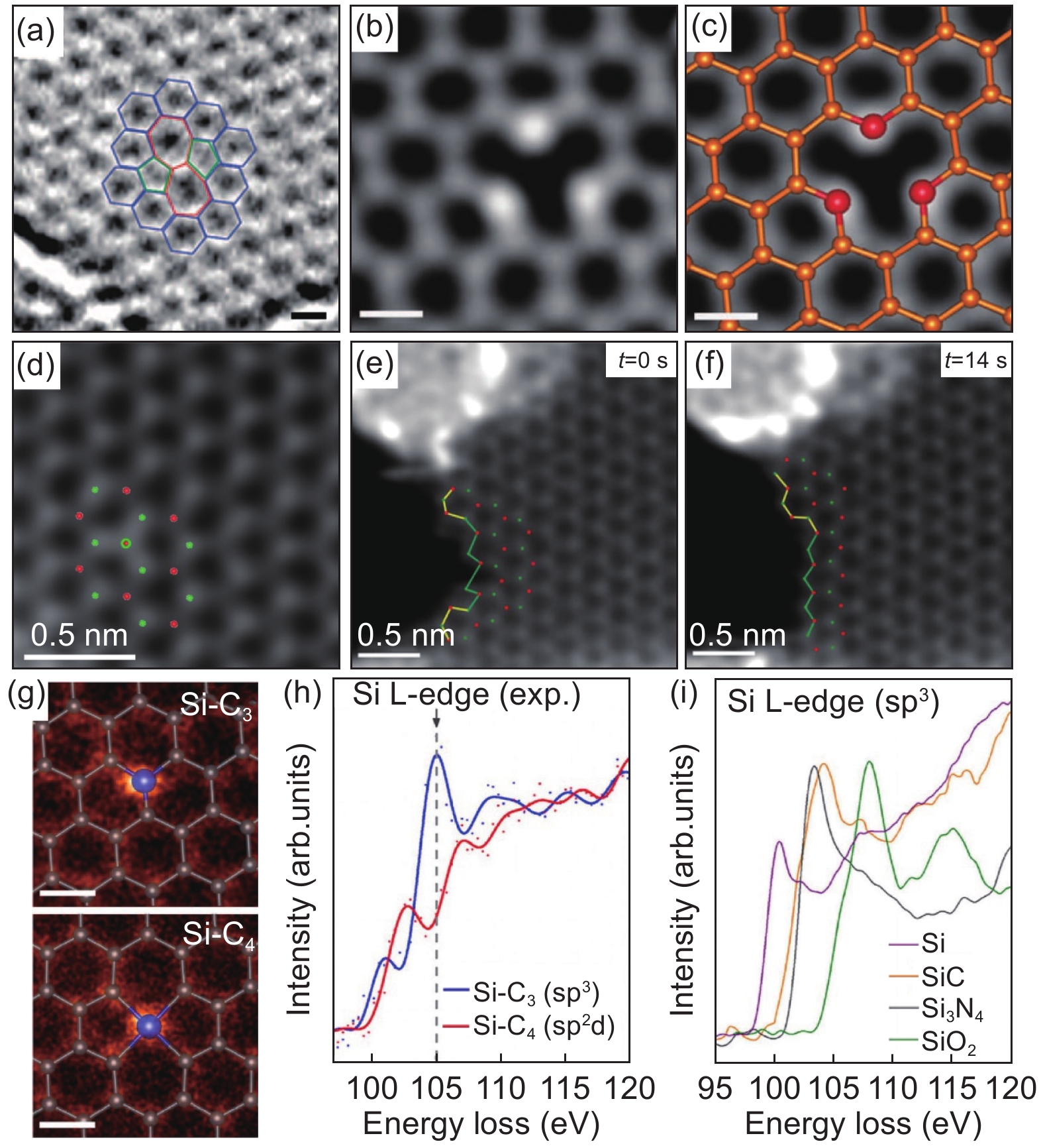
Figure 6. Atomic structures of defects in graphene and alike 2D materials. (a) HRTEM image of SW defect in graphene (Scale bar is 0.2 nm)[3]. (b-c) STEM-ADF image of 3 oxygen atoms in graphene multivacancies (Scale bar is 0.2 nm)[10]. (d-f) STEM-ADF image of a nitrogen anti-site NB in a suspended h-BN monolayer, and its edge reconstruction[11] (Red and green dots correspond to B and N atom). (g-i) STEM-ADF image and ELNES for threefold and fourfold coordinated Si impurities in monolayer graphene (Scale bar is 0.2 nm)[51].

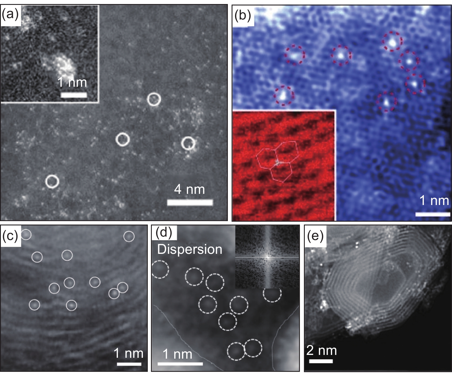
Figure 7. STEM characterization of carbon-supported SACs. (a) ADF image of Pt/graphene catalyst for methanol oxidation[55]. (b) HAADF image of single Ni doped graphene for HER[56]. (c) ADF image of Nb atoms anchored in onion-like carbon shells used for ORR catalysis[9]. (d) ADF image of dispersed single W atoms in high-defective graphene layers of carbon onions for ORR catalysis[59]. (f) ADF image Pt atoms in carbon onions, which is catalytically active for chemoselective hydrogenation of nitroarenes[60].
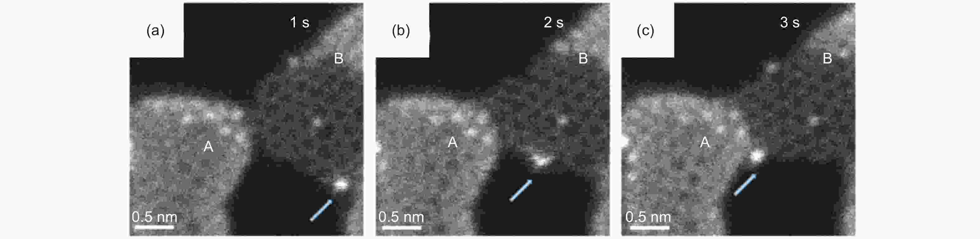
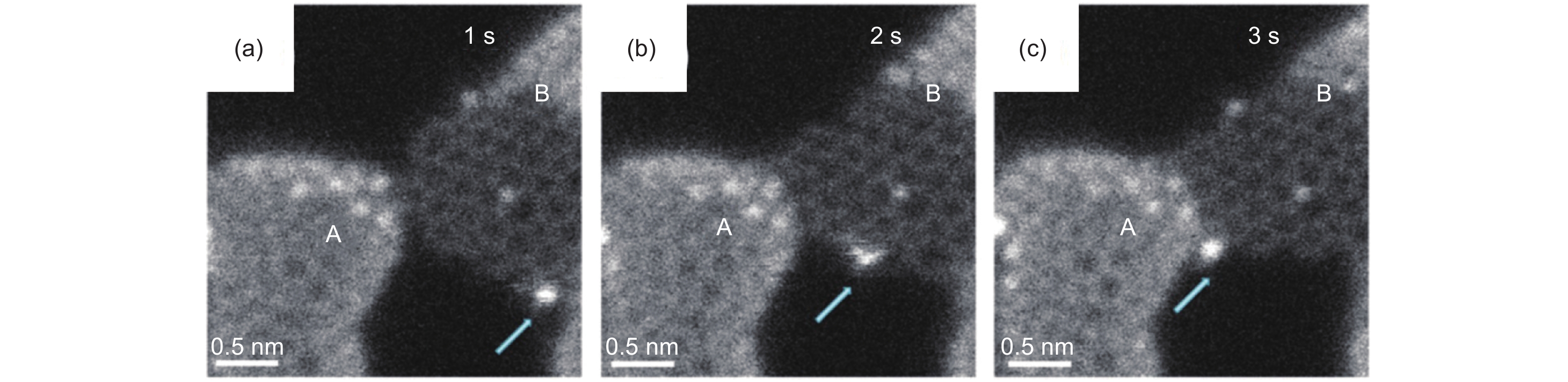
Figure 8. Dynamic structure evolutions of graphene-based materials. (a-c) STEM-ADF images show dynamics of a Ni atom at t = 1 s, 2 s, and 3 s in a graphene nanomesh, respectively[65].

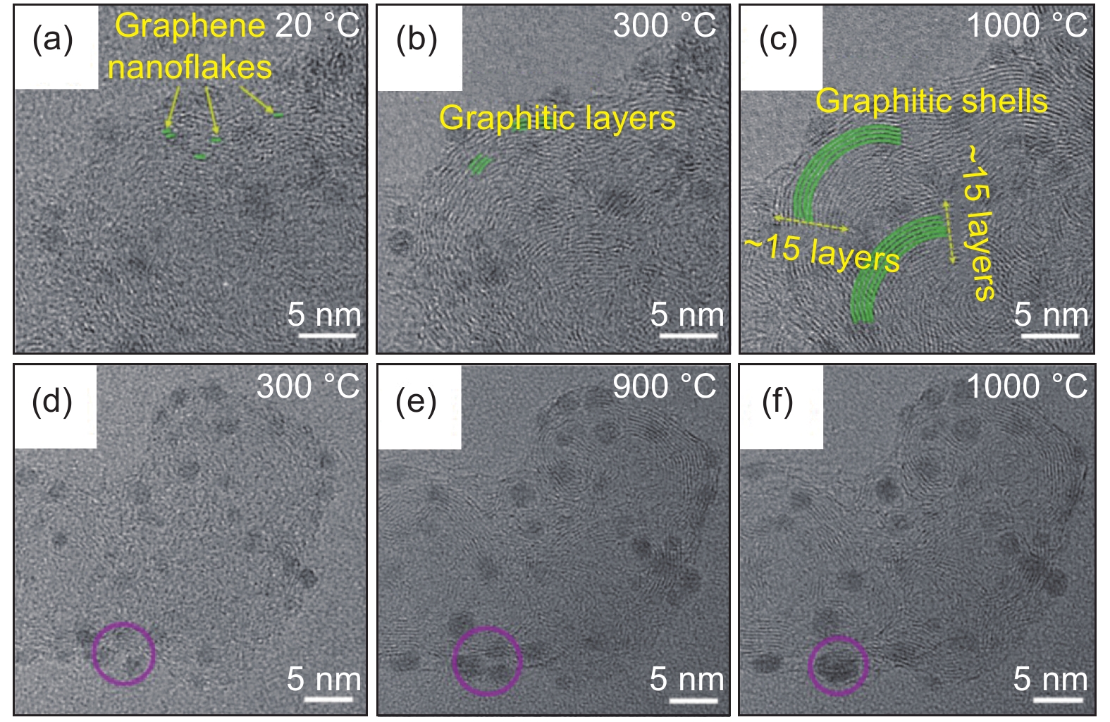
Figure 9. HRTEM image series demonstrate structure evolutions of (a-c) graphitic shells and (d-f) aggregation of Ru nanoparticles in Ru@GNs catalyst during in situ heating[68].
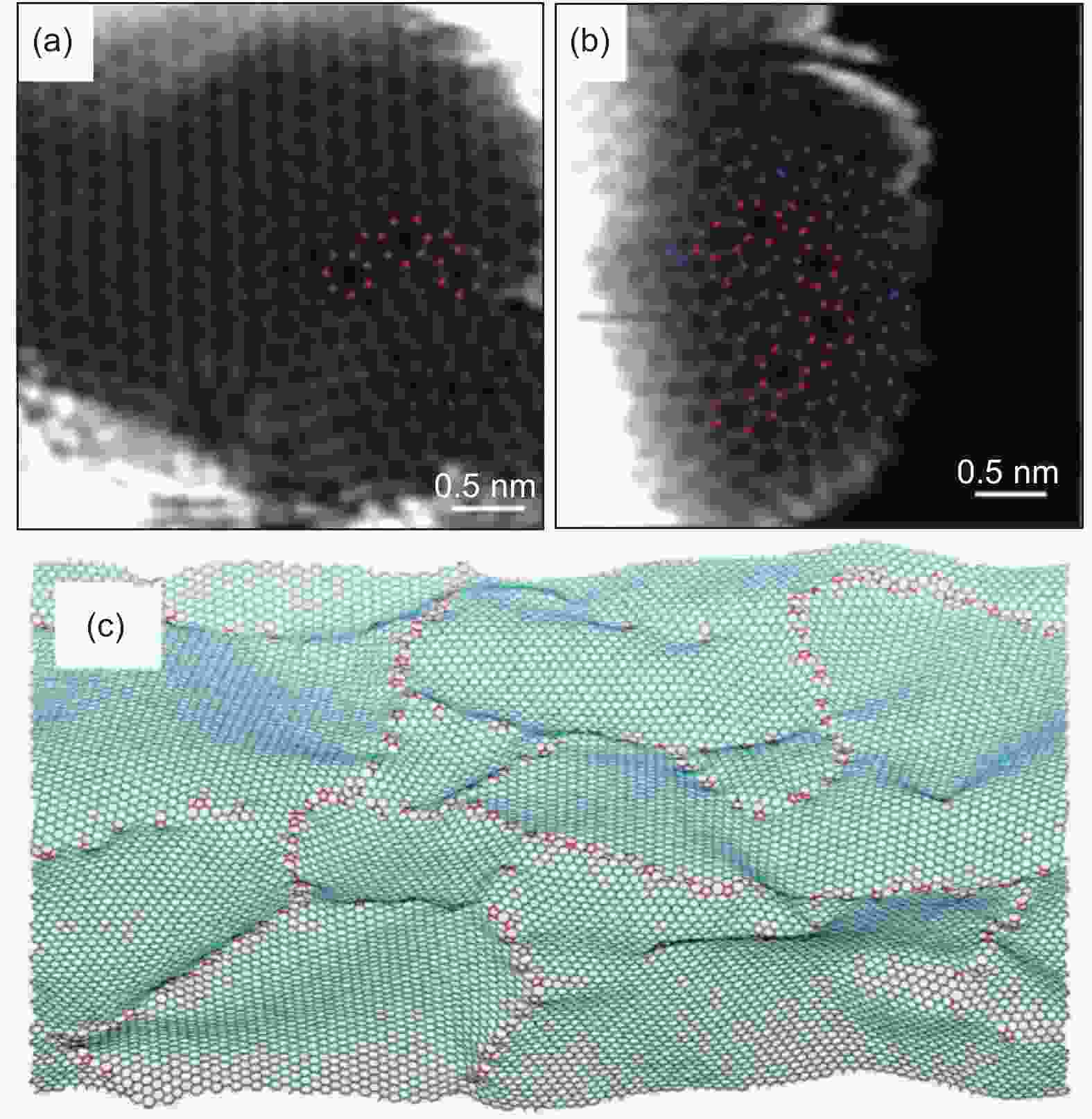
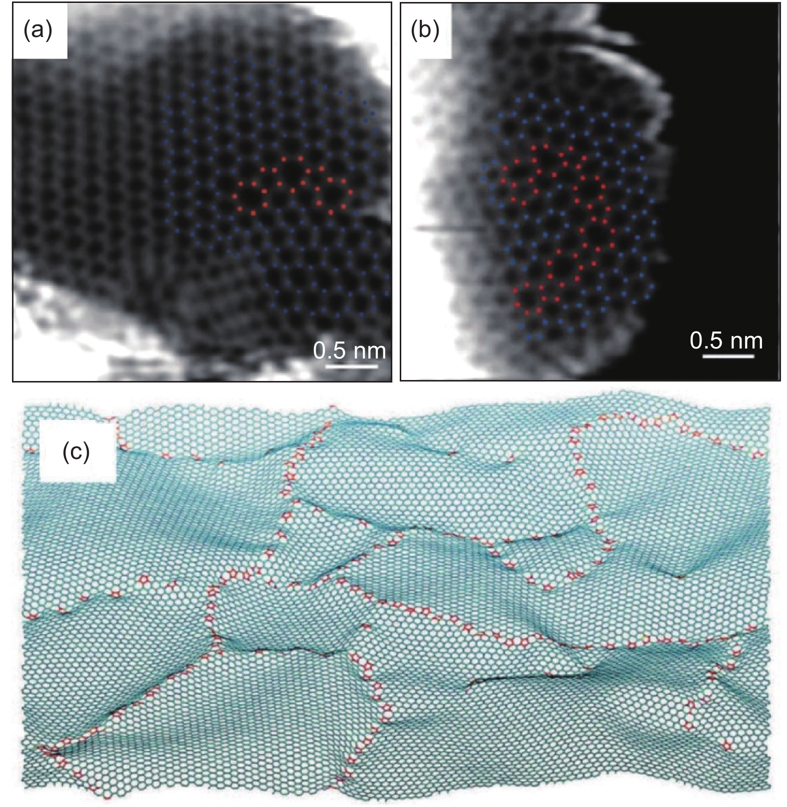
Figure 10. (a, b) ADF STEM images of a wood-based nanoporous carbon with large areas of hexagonal lattice (marked in blue) and a few five- and seven-atom ring defects (marked in red). (c) Segment of a simulated defective graphene sheet, with five to seven dislocation structures arranged similarly to microscopy observations.
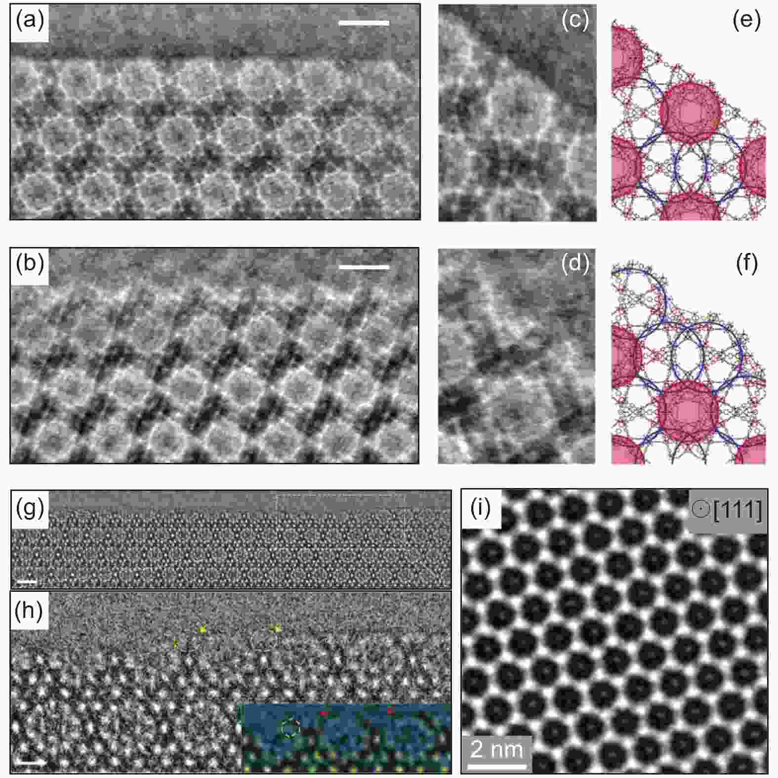
Figure 11. Characterization of MOFs with low dose electron microscopy. (a, b) iDPC-STEM image of MIL-101 (111) surface with different termination cages (Scale bars: 5 nm). (c, d) The structures of single unit cells at the two types of surface terminations (Scale bars: 3 nm). (e, f) The structural models show the (111) surfaces terminated by different cages[20]. (g) HRTEM image of sublayer (
$ \bar{1}11 $ ) surface of MIL-101 (Scale bar: 5 nm). (h) Magnified image showing the evolution from sublayer to stable (111) surface (Scale bar: 2 nm).[19](i) Cryo-TEM image of CO2-filled ZIF-8 particle along the <111> projection, with Zn clusters and adsorbed CO2 molecules being identified.[74] In the referred work above, total electron dose for iDPC-STEM is 40 e-Å–2, and electron dose for low dose TEM is usually less than 10.4 e-Å–2 accumulated from tens of dose fractionation frames.
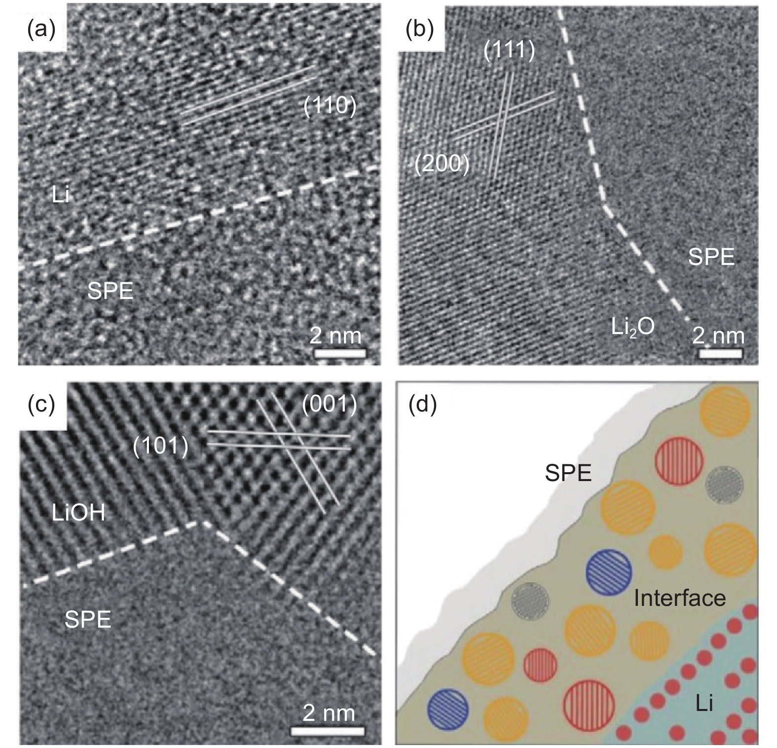
Figure 12. Atomic resolution cryo-TEM of SEI. (a-c) HRTEM images of the interface between crystalline Li, Li2O, LiOH and amorphous SPE. (d) Schematic diagram for the identified mosaic structure of SEI.
-
[1] Novoselov K S, Geim A K, Morozov S V, et al. Electric field effect in atomically thin carbon films[J]. Science,2004,306(5696):666-669. doi: 10.1126/science.1102896 [2] Meyer J C, Geim A K, Katsnelson M I, et al. The structure of suspended graphene sheets[J]. Nature,2007,446(7131):60-63. doi: 10.1038/nature05545 [3] Meyer J C, Kisielowski C, Erni R, et al. Direct imaging of lattice atoms and topological defects in graphene membranes[J]. Nano Letters,2008,8(11):3582-3586. doi: 10.1021/nl801386m [4] Gass M H, Bangert U, Bleloch A L, et al. Free-standing graphene at atomic resolution[J]. Nature Nanotechnology,2008,3(11):676-681. doi: 10.1038/nnano.2008.280 [5] Suenaga K, Koshino M. Atom-by-atom spectroscopy at graphene edge[J]. Nature,2010,468(7327):1088-1090. doi: 10.1038/nature09664 [6] Krivanek O L, Chisholm M F, Nicolosi V, et al. Atom-by-atom structural and chemical analysis by annular dark-field electron microscopy[J]. Nature,2010,464(7288):571-574. doi: 10.1038/nature08879 [7] Zhang X, Guo J, Guan P, et al. Gigahertz dielectric polarization of substitutional single niobium atoms in defective graphitic layers[J]. Physical Review Letters,2015,115(14):147601. doi: 10.1103/PhysRevLett.115.147601 [8] Geim A K, Novoselov K S. The rise of graphene[J]. Nature Materials,2007,6(3):183-191. doi: 10.1038/nmat1849 [9] Zhang X, Guo J, Guan P, et al. Catalytically active single-atom niobium in graphitic layers[J]. Nature Communications,2013,4(1):1924. doi: 10.1038/ncomms2929 [10] Guo J, Lee J, Contescu C I, et al. Crown ethers in graphene[J]. Nature Communications,2014,5(1):5389. doi: 10.1038/ncomms6389 [11] Liu P, Tian H, Windl W, et al. Direct imaging of the nitrogen-rich edge in monolayer hexagonal boron nitride and its band structure tuning[J]. Nanoscale,2019,11(43):20676-20684. doi: 10.1039/C9NR07147D [12] Zhang Z, Liu W, Zhang B, et al. Defect-nucleated phase transition in atomically-thin WS2[J]. 2D Materials,2021,8(2):025017. doi: 10.1088/2053-1583/abd6b4 [13] Song Y, Xu B, Liao T, et al. Electronic structure tuning of 2D metal (hydr)oxides nanosheets for electrocatalysis[J]. Small,2021,17(9):2002240. doi: 10.1002/smll.202002240 [14] Wang J, Liao T, Wei Z, et al. Heteroatom-doping of non-noble metal-based catalysts for electrocatalytic hydrogen evolution: an electronic structure tuning strategy[J]. Small Methods,2021,n/a(n/a):2000988. [15] Erni R, Rossell M D, Kisielowski C, et al. Atomic-resolution imaging with a sub-50-pm electron probe[J]. Physical Review Letters,2009,102(9):096101. doi: 10.1103/PhysRevLett.102.096101 [16] Egerton R F, Li P, Malac M. Radiation damage in the TEM and SEM[J]. Micron,2004,35(6):399-409. doi: 10.1016/j.micron.2004.02.003 [17] Egerton R F. Mechanisms of radiation damage in beam-sensitive specimens, for TEM accelerating voltages between 10 and 300 kV[J]. Microscopy Research and Technique,2012,75(11):1550-1556. doi: 10.1002/jemt.22099 [18] Zhu Y, Ciston J, Zheng B, et al. Unravelling surface and interfacial structures of a metal–organic framework by transmission electron microscopy[J]. Nature Materials,2017,16(5):532-536. doi: 10.1038/nmat4852 [19] Han X, Liu P, Lin F, et al. Structures and structural evolution of sublayer surfaces of metal–organic frameworks[J]. Angewandte Chemie International Edition,2020,59(48):21419-21424. doi: 10.1002/anie.202008100 [20] Shen B, Chen X, Shen K, et al. Imaging the node-linker coordination in the bulk and local structures of metal-organic frameworks[J]. Nature Communications,2020,11(1):2692. doi: 10.1038/s41467-020-16531-y [21] Li Y, Li Y, Pei A, et al. Atomic structure of sensitive battery materials and interfaces revealed by cryo–electron microscopy[J]. Science,2017,358(6362):506. doi: 10.1126/science.aam6014 [22] Callaway E. 'It opens up a whole new universe': revolutionary microscopy technique sees individual atoms for first time[J]. Nature,2020,582(7811):156-157. doi: 10.1038/d41586-020-01658-1 [23] NobelPrize.org. The Nobel Prize in Physics 1986[EB/OL]. <https://www.nobelprize.org/prizes/physics/1986/summary/>. [24] Hirsch P B, Horne R W, Whelan M J. Direct observations of the arrangement and motion of dislocations in aluminium[J]. Philosophical Magazine,1956,86(29-31):4553-4572. [25] Shechtman D, Blech I, Gratias D, et al. Metallic phase with long-range orientational order and no translational symmetry[J]. Physical Review Letters,1984,53(20):1951-1953. doi: 10.1103/PhysRevLett.53.1951 [26] Zhang Z, Ye H, Kuo K. A new icosahedral phase with m35 symmetry[J]. Philosophical Magazine A,1985,52(6):L49-L52. doi: 10.1080/01418618508242135 [27] Jiang W, Hei K, Guo Y, et al. Tenfold twins in a rapidly quenched NiZr alloy[J]. Philosophical Magazine A,1985,52(6):L53-L58. doi: 10.1080/01418618508242136 [28] Iijima S. Helical microtubules of graphitic carbon[J]. Nature,1991,354(6348):56-58. doi: 10.1038/354056a0 [29] Shang T, Liu X, Gu L. Interface of transition metal oxides at the atomic scale[J]. Science China Physics, Mechanics & Astronomy,2016,59(9):697001. [30] Crewe A V, Wall J, Langmore J. Visibility of single atoms[J]. Science,1970,168(3937):1338. doi: 10.1126/science.168.3937.1338 [31] Pennycook S J, Boatner L A. Chemically sensitive structure-imaging with a scanning transmission electron microscope[J]. Nature,1988,336(6199):565-567. doi: 10.1038/336565a0 [32] Pennycook S J, Jesson D E. High-resolution incoherent imaging of crystals[J]. Physical Review Letters,1990,64(8):938-941. doi: 10.1103/PhysRevLett.64.938 [33] Zhang Q, Xiao D, Gu L. Aberration-corrected scanning transmission electron microscopy for complex transition metal oxides[J]. Chin. Phys. B,2016,25(6):66803-066803. doi: 10.1088/1674-1056/25/6/066803 [34] Geuens P, Dyck D V. The s‐state model for electron channeling in high‐resolution electron microscopy[J]. Advances in Imaging and Electron Physics: Elsevier,2005:111-226. [35] Wen Y, Shang T, Gu L. Analytical ABF-STEM imaging of Li ions in rechargeable batteries[J]. Microscopy,2017,66(1):25-38. [36] Ishikawa R, Okunishi E, Sawada H, et al. Direct imaging of hydrogen-atom columns in a crystal by annular bright-field electron microscopy[J]. Nature Materials,2011,10(4):278-281. doi: 10.1038/nmat2957 [37] Findlay S D, Kohno Y, Cardamone L A, et al. Enhanced light element imaging in atomic resolution scanning transmission electron microscopy[J]. Ultramicroscopy,2014,136:31-41. doi: 10.1016/j.ultramic.2013.07.019 [38] Scherzer O. The theoretical resolution limit of the electron microscope[J]. Journal of Applied Physics,1949,20(1):20-29. doi: 10.1063/1.1698233 [39] Bleloch A, Lupini A. Imaging at the picoscale[J]. Materials Today,2004,7(12):42-48. [40] Haider M, Uhlemann S, Schwan E, et al. Electron microscopy image enhanced[J]. Nature,1998,392(6678):768-769. doi: 10.1038/33823 [41] Batson P E, Dellby N, Krivanek O L. Sub-ångstrom resolution using aberration corrected electron optics[J]. Nature,2002,418(6898):617-620. doi: 10.1038/nature00972 [42] Nellist P D, Chisholm M F, Dellby N, et al. Direct sub-angstrom imaging of a crystal lattice[J]. Science,2004,305(5691):1741. doi: 10.1126/science.1100965 [43] wolffund.org. Wolf Prize in Physics 2011[EB/OL]. <https://wolffund.org.il/2018/12/11/harald-rose/>. [44] kavliprize.org. 2020 Kavli Prize in Nanoscience[EB/OL]. <http://kavliprize.org/prizes-and-laureates/prizes/2020-kavli-prize-nanoscience#page-title>. [45] Partoens B, Peeters F M. From graphene to graphite: electronic structure around the K point[J]. Physical Review B,2006,74(7):075404. doi: 10.1103/PhysRevB.74.075404 [46] Castro Neto A H, Guinea F, Peres N M R, et al. The electronic properties of graphene[J]. Reviews of Modern Physics,2009,81(1):109-162. doi: 10.1103/RevModPhys.81.109 [47] NobelPrize.org. The Nobel Prize in Physics 2010[EB/OL]. <https://www.nobelprize.org/prizes/physics/2010/summary/>. [48] Zobelli A, Gloter A, Ewels C P, et al. Electron knock-on cross section of carbon and boron nitride nanotubes[J]. Phys. Rev. B,2007,75(24):245402. doi: 10.1103/PhysRevB.75.245402 [49] Huang P Y, Ruiz-Vargas C S, van der Zande A M, et al. Grains and grain boundaries in single-layer graphene atomic patchwork quilts[J]. Nature,2011,469(7330):389-392. doi: 10.1038/nature09718 [50] Liu P, Guo J, Liu L, et al. Direct observation of defects in hexagonal boron nitride monolayers[C]. Microscopy and Microanalysis, 2014 20 (S3): 1738-1740. [51] Zhou W, Kapetanakis M D, Prange M P, et al. Direct determination of the chemical bonding of individual impurities in graphene[J]. Physical Review Letters,2012,109(20):206803. doi: 10.1103/PhysRevLett.109.206803 [52] Toh C, Zhang H, Lin J, et al. Synthesis and properties of free-standing monolayer amorphous carbon[J]. Nature,2020,577(7789):199-203. doi: 10.1038/s41586-019-1871-2 [53] Yang X, Wang A, Qiao B, et al. Single-atom catalysts: a new frontier in heterogeneous catalysis[J]. Accounts of Chemical Research,2013,46(8):1740-1748. doi: 10.1021/ar300361m [54] Li H, Zhang H, Yan X, et al. Carbon-supported metal single atom catalysts[J]. New Carbon Materials,2018,33(1):1-11. doi: 10.1016/S1872-5805(18)60322-1 [55] Sun S, Zhang G, Gauquelin N, et al. Single-atom catalysis using Pt/graphene achieved through atomic layer deposition[J]. Scientific Reports,2013,3(1):1775. doi: 10.1038/srep01775 [56] Qiu H, Ito Y, Cong W, et al. Nanoporous graphene with single-atom nickel dopants: an efficient and stable catalyst for electrochemical hydrogen production[J]. Angewandte Chemie International Edition,2015,54(47):14031-14035. doi: 10.1002/anie.201507381 [57] Fei H, Dong J, Arellano-Jiménez M J, et al. Atomic cobalt on nitrogen-doped graphene for hydrogen generation[J]. Nature Communications,2015,6(1):8668. doi: 10.1038/ncomms9668 [58] Fei H, Dong J, Feng Y, et al. General synthesis and definitive structural identification of MN4C4 single-atom catalysts with tunable electrocatalytic activities[J]. Nature Catalysis,2018,1(1):63-72. doi: 10.1038/s41929-017-0008-y [59] Guo J, Mao Z, Yan X, et al. Ultrasmall tungsten carbide catalysts stabilized in graphitic layers for high-performance oxygen reduction reaction[J]. Nano Energy,2016,28:261-268. doi: 10.1016/j.nanoen.2016.08.045 [60] Yan X, Duan P, Zhang F, et al. Stable single-atom platinum catalyst trapped in carbon onion graphitic shells for improved chemoselective hydrogenation of nitroarenes[J]. Carbon,2019,143:378-384. doi: 10.1016/j.carbon.2018.11.021 [61] Zhang X, Rao Y, Guo J, et al. Multiple-phase carbon-coated FeSn2/Sn nanocomposites for high-frequency microwave absorption[J]. Carbon,2016,96:972-979. doi: 10.1016/j.carbon.2015.09.087 [62] Warner J, Rümmeli M, Ge L, et al. Structural transformations in graphene studied with high spatial and temporal resolution[J]. Nature Nanotechnology,2009,4(8):500-504. doi: 10.1038/nnano.2009.194 [63] Girit Ç Ö, Meyer J C, Erni R, et al. Graphene at the edge: stability and dynamics[J]. Science,2009,323(5922):1705. doi: 10.1126/science.1166999 [64] Warner J H, Margine E R, Mukai M, et al. Dislocation-driven deformations in graphene[J]. Science,2012,337(6091):209. doi: 10.1126/science.1217529 [65] Zhang H, Liu W, Zhang Z, et al. Direct imaging of a single Ni atom cutting graphene to form a graphene nanomesh[J]. Physical Chemistry Chemical Physics,2018,20(42):26814-26818. doi: 10.1039/C8CP03711F [66] Luo C, Wang C L, Wu X, Zhang J, Chu J H. In situ transmission electron microscopy characterization and manipulation of two-dimensional layered materials beyond graphene[J]. Small, 2017, 13(35). https://doi.org/10.1002/smll.201604259. [67] Su Q, Du G, Guo J, et al. Recent progress of in situ transmission electron microscopy on electrochemical energy storage[J]. Materials China, 2020, 39. [68] Shen Y, Liu P, Du J, et al. Defect engineering in graphene-based nanospheres enhanced hydrogen evolution reaction performance of ruthenium nanoparticles[J]. Carbon,2020,166:388-395. doi: 10.1016/j.carbon.2020.05.033 [69] Meng Y, Young T M, Liu P, et al. Ultralight carbon aerogel from nanocellulose as a highly selective oil absorption material[J]. Cellulose,2015,22(1):435-447. doi: 10.1007/s10570-014-0519-5 [70] Meng Y, Contescu C I, Liu P, et al. Understanding the local structure of disordered carbons from cellulose and lignin[J]. Wood Science and Technology,2021,55:587-606. doi: 10.1007/s00226-021-01286-6 [71] Pei L, Cao H, Yang L, et al. Hard carbon derived from waste tea biomass as high-performance anode material for sodium-ion batteries[J]. Ionics,2020,26(11):5535-5542. doi: 10.1007/s11581-020-03723-1 [72] Pei L, Yang L, Cao H, et al. Cost-effective and renewable paper derived hard carbon microfibers as superior anode for sodium-ion batteries[J]. Electrochimica Acta,2020,364:137313. doi: 10.1016/j.electacta.2020.137313 [73] Guo J, Morris J R, Ihm Y, et al. Topological defects: origin of nanopores and enhanced adsorption performance in nanoporous carbon[J]. Small,2012,8(21):3283-3288. doi: 10.1002/smll.201200894 [74] Li Y, Wang K, Zhou W, et al. Cryo-EM structures of atomic surfaces and host-guest chemistry in metal-organic frameworks[J]. Matter,2019,1(2):428-438. doi: 10.1016/j.matt.2019.06.001 [75] Lazić I, Bosch E G T, Lazar S. Phase contrast STEM for thin samples: Integrated differential phase contrast[J]. Ultramicroscopy,2016,160:265-280. doi: 10.1016/j.ultramic.2015.10.011 [76] Ren X, Zhang X, Xu R, et al. Analyzing energy materials by cryogenic electron microscopy[J]. Advanced Materials,2020,32(24):1908293. doi: 10.1002/adma.201908293 [77] Liu Y, Ju Z, Zhang B, Wang Y, Nai J, Liu T, Tao X. Visualizing the sensitive lithium with atomic precision: cryogenic electron microscopy for batteries[J]. Accounts of Chemical Research, 2021, 54(9): 2088–2099. [78] Sheng O, Zheng J, Ju Z, et al. In situ vonstruction of a LiF-rnriched interface for stable all-solid-state batteries and its origin revealed by cryo-TEM[J]. Advanced Materials,2020,32(34):2000223. doi: 10.1002/adma.202000223 [79] NobelPrize.org. The Nobel Prize in Chemistry 2017[EB/OL]. <https://www.nobelprize.org/prizes/chemistry/2017/summary/>. [80] Yao H, Song Y, Chen Y, et al. Molecular Architecture of the SARS-CoV-2 Virus[J]. Cell,2020,183(3):730-738. doi: 10.1016/j.cell.2020.09.018 [81] Bai R, Wan R, Yan C, et al. Mechanism of spliceosome remodeling by the ATPase/helicase Prp2 and its coactivator Spp2[J]. Science,2021,371(6525):eabe8863. doi: 10.1126/science.abe8863 -


 下载:
下载:


 点击查看大图
计量
- 文章访问数: 2285
- HTML全文浏览量: 1319
- PDF下载量: 204
- 被引次数: 0