-
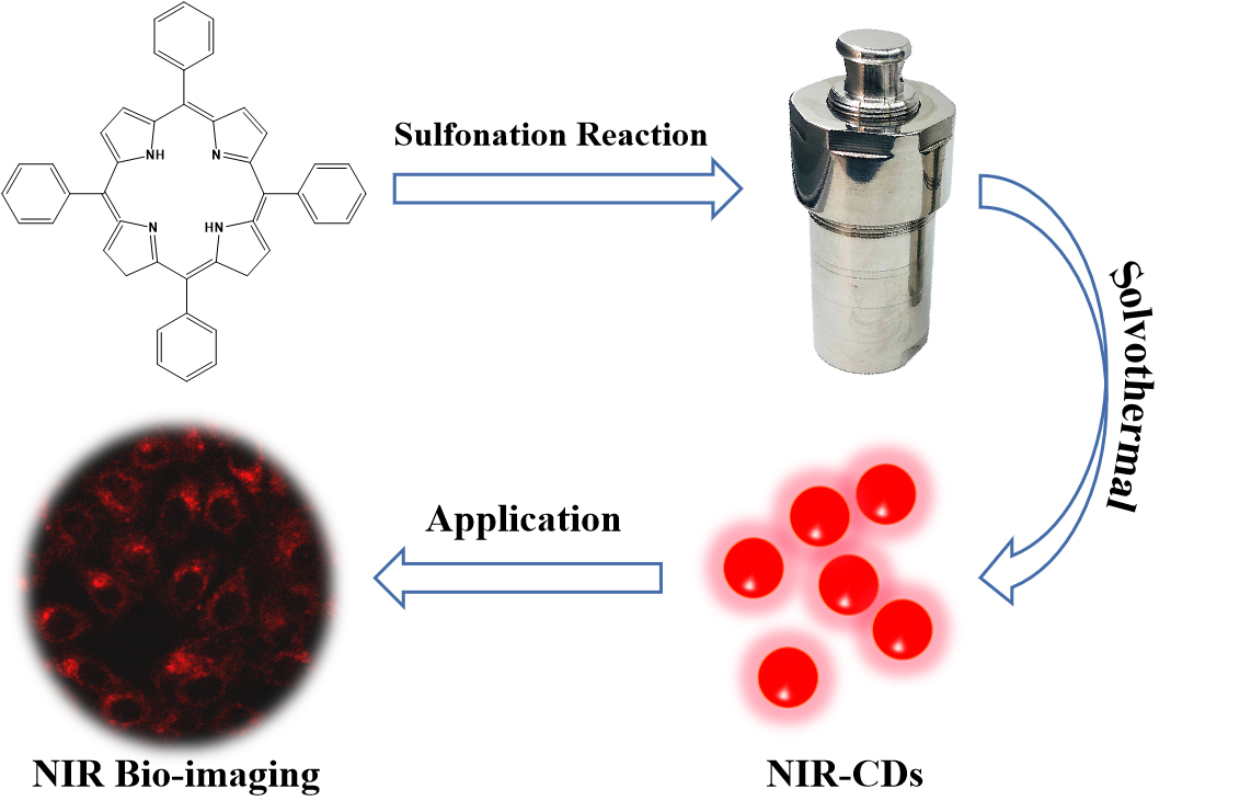
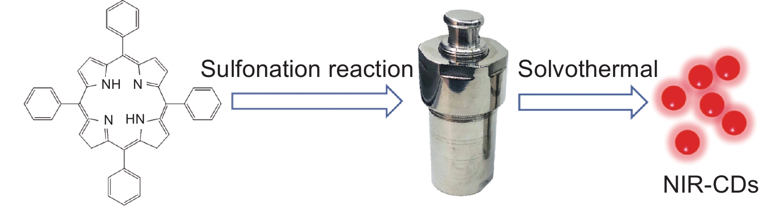
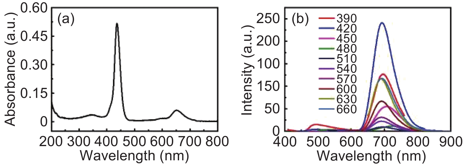
摘要: 由于红光/近红外发射具有深层组织穿透力强、自体荧光小、对生物组织损伤小等特点,具有上述特性的碳点的制备与生物成像应用备受关注。本文以磺化四苯基卟啉为前驱体,采用溶剂热法合成近红外发射的荧光碳点(NIR-CDs)。NIR-CDs的最大发射峰位于692 nm,其荧光发射具有激发波长非依赖性,经分析NIR-CDs的近红外荧光发射主要源于分子态发光。此外,NIR-CDs还具有良好的水溶性和生物相容性、丰富的表面官能团、低毒性和优异的细胞标记能力,证实了NIR-CDs在细胞近红外成像中的应用潜力。本研究有望促进面向生物应用的近红外荧光碳点的发展,推动新型碳点的研究与实际应用。Abstract: It is very difficult to prepare red/near-infrared emission carbon dots (CDs) for bio-imaging applications which are needed because of their deep tissue penetration, minimal auto-fluorescence, and low emission light damage to bio-tissues. Near-infrared emitting CDs (NIR-CDs) were synthesized from sulfonated tetraphenylporphyrin using a solvothermal method. They have excitation-independent properties with a maximum emission at 692 nm. Studies showed that this unique near-infrared emission mainly originated from the aggregated molecular states of the CDs. The NIR-CDs showed good water solubility, exceptional biocompatibility, low toxicity, and superior cellular labelling ability. This work could significantly advance the structural design and preparation of NIR-CDs and corresponding bio-imaging applications.
-
Key words:
- Near-infrared emission /
- Molecular states /
- Cellular labelling /
- Bio-imaging
-
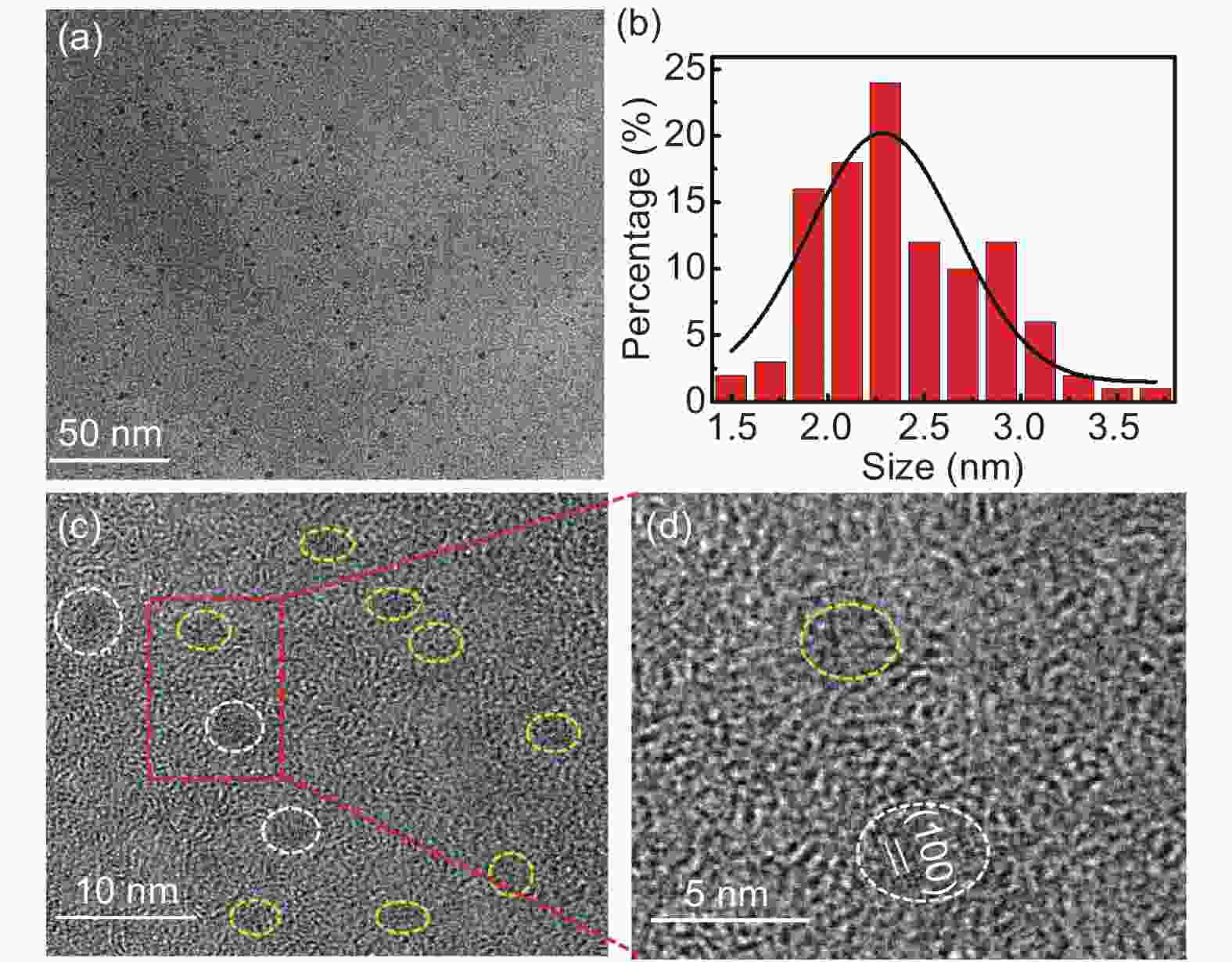
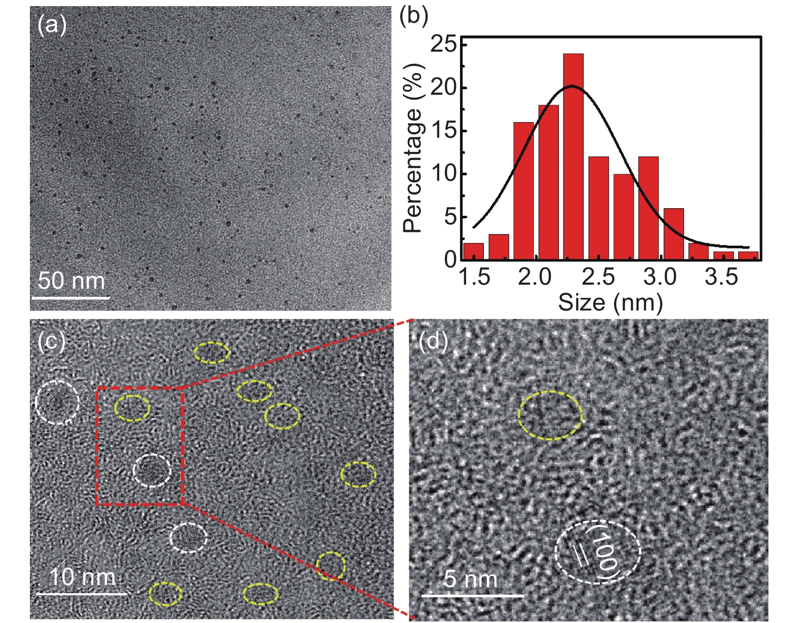
Figure 2. (a) TEM and (b) the size distribution histogram of the NIR-CDs; (c, d) HRTEM images of the NIR-CDs. The white circles stand for crystalline NIR-CDs, and yellow circles represent amorphous CDs.
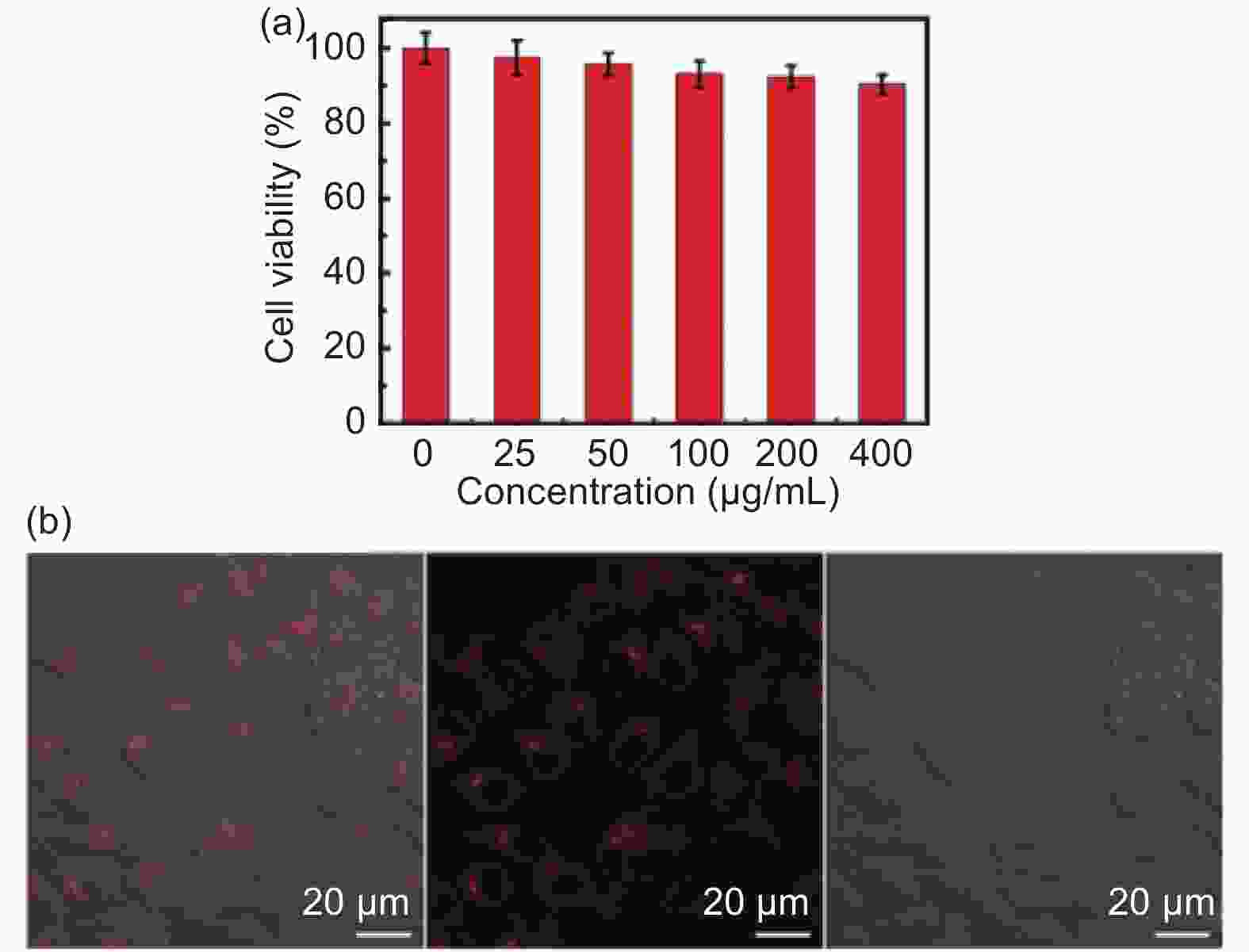
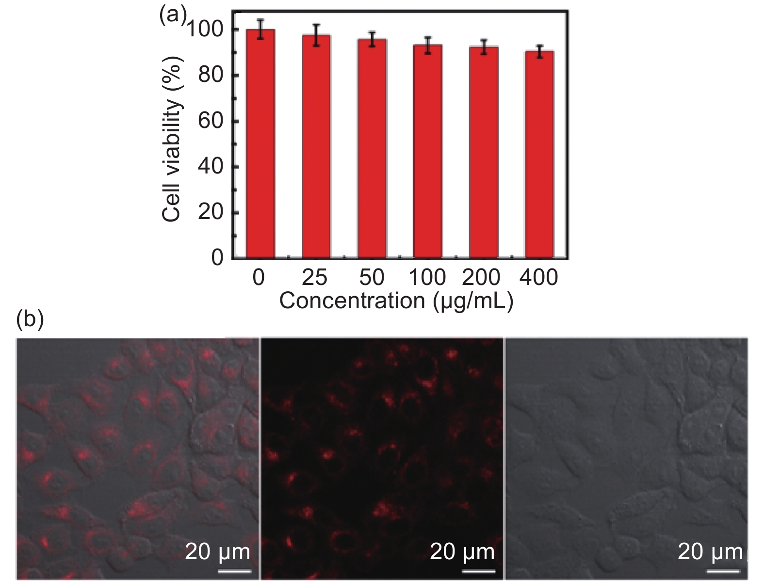
Figure 6. (a) Cell viability of HeLa cells after incubation with various concentrations of NIR-CDs; (b) Confocal images of Hela cells incubated with NIR-CDs at the concentration of 200 µg mL−1 obtained under dark field (right image), bright field (middle image) and their merged (left) image at the excitation of 543 nm laser.
-
[1] Li L P, Lu C C, Li S J, et al. A high-yield and versatile method for the synthesis of carbon dots for bioimaging applications[J]. Journal of Materials Chemistry B,2017,5:1935-1942. [2] Jia Q Y, Zhao Z Y, Liang K, et al. Recent advances and prospects of carbon dots in cancer nanotheranostics[J]. Materials Chemistry Frontiers,2020,4:449-471. doi: 10.1039/C9QM00667B [3] Yang S T, Cao L, Luo P G, et al. Carbon dots for optical imaging in vivo[J]. Journal of the American Chemical Society,2009,131:11308-11309. doi: 10.1021/ja904843x [4] Li J Y, Liu Y, Shu Q W, et al. One-pot hydrothermal synthesis of carbon dots with efficient up- and down-converted photoluminescence for the sensitive detection of morin in a dual-readout assay[J]. Langmuir,2017,33:1043-1050. doi: 10.1021/acs.langmuir.6b04225 [5] Reyes D, Camacho M, Camacho M, et al. Laser ablated carbon nanodots for light emission[J]. Nanoscale Research Letters,2016,11:424. doi: 10.1186/s11671-016-1638-8 [6] De Medeiros T V, Manioudakis J, Noun F, et al. Microwave-assisted synthesis of carbon dots and their applications[J]. Journal of Materials Chemistry C,2019,7:7175-7195. doi: 10.1039/C9TC01640F [7] Liu M L, Xu Y H, NIU F, et al. Carbon quantum dots directly generated from electrochemical oxidation of graphite electrodes in alkaline alcohols and the applications for specific ferric ion detection and cell imaging[J]. Analyst,2016,141(9):2657-2664. doi: 10.1039/C5AN02231B [8] Zhang X Y, Jiang M Y, Niu N, et al. Natural-product-derived carbon dots: from natural products to functional materials[J]. ChemSusChem,2018,11:11-24. doi: 10.1002/cssc.201701847 [9] Jiang K, Zhang L, Lu J, et al. Triple-mode emission of carbon dots: Applications for advanced anti-counterfeiting[J]. Angewandte Chemie International Edition,2016,55:7231-7235. doi: 10.1002/anie.201602445 [10] Wu Z L, Zhang P, Gao M X, et al. One-pot hydrothermal synthesis of highly luminescent nitrogen-doped amphoteric carbon dots for bioimaging from Bombyx mori silk-natural proteins[J]. Journal of Materials Chemistry B,2013,1:2868-2873. doi: 10.1039/c3tb20418a [11] Unnikrishnan B, Wu R S, Wei S C, et al. Fluorescent carbon dots for selective labelling of subcellular organelles[J]. ACS Omega,2020,5:11248-11261. doi: 10.1021/acsomega.9b04301 [12] Du J J, Xu N, Fan J L, et al. Carbon dots for in vivo bioimaging and theranostics[J]. Small,2019,15:1805087. doi: 10.1002/smll.201805087 [13] Zhang J, Yu S H. Carbon dots: large-scale synthesis, sensing and bioimaging[J]. Materials Today,2016,19:382-393. doi: 10.1016/j.mattod.2015.11.008 [14] Wang Q L, Huang X X, Long Y J, et al. Hollow luminescent carbon dots for drug delivery[J]. Carbon,2013,59:192-199. doi: 10.1016/j.carbon.2013.03.009 [15] Sun S, Zhang L, Jiang K, et al. Toward high-efficient red emissive carbon dots: Facile preparation, unique properties, and applications as multifunctional theranostic agents[J]. Chemistry of Materials,2016,28:8659-8668. doi: 10.1021/acs.chemmater.6b03695 [16] Li H X, Su D D, Gao H, et al. Design of red emissive carbon dots: Robust performance for analytical applications in pesticide monitoring[J]. Analytical Chemistry,2020,92:3198-3205. doi: 10.1021/acs.analchem.9b04917 [17] Gao W L, Song H H, Wang X, et al. Carbon dots with red emission for sensing of Pt2+, Au3+, and Pd2+ and their bioapplications in vitro and in vivo[J]. ACS Applied Materials & Interfaces,2018,10:1147-1154. [18] Gao Y F, Jiao Y, Lu W J, et al. Carbon dots with red emission as a fluorescent and colourimeteric dual-readout probe for the detection of chromium(vi) and cysteine and its logic gate operation[J]. Journal of Materials Chemistry B,2018,6:6099-6107. doi: 10.1039/C8TB01580E [19] Jiang K, Sun S, Zhang L, et al. Red, green, and blue luminescence by carbon dots: full-colour emission tuning and multicolour cellular imaging[J]. Angewandte Chemie International Edition,2015,54:5360-5363. doi: 10.1002/anie.201501193 [20] Tan X Y, Li Y C, Li X H, et al. Electrochemical synthesis of small-sized red fluorescent graphene quantum dots as a bioimaging platform[J]. Chemical Communications,2015,51:2544-2546. doi: 10.1039/C4CC09332A [21] Liu M L, Chen B B, Li C M, et al. Carbon dots: Synthesis, formation mechanism, fluorescence origin and sensing application[J]. Green Chemistry,2019,21:449-471. doi: 10.1039/C8GC02736F [22] Li L P, Zhang R P, Lu C X, et al. In situ synthesis of NIR-light emitting carbon dots derived from spinach for bio-imaging applications[J]. Journal of Materials Chemistry B,2017,5:7328-7334. doi: 10.1039/C7TB00634A [23] Pan L L, Sun S, Zhang L, et al. Near-infrared emissive carbon dots for two-photon fluorescence bioimaging[J]. Nanoscale,2016,8:17350-17356. doi: 10.1039/C6NR05878G [24] Jiang K, Hu S Z, Wang Y C, et al. Photo-stimulated polychromatic room temperature phosphorescence of carbon dots[J]. Small,2020,16:2001909. doi: 10.1002/smll.202001909 [25] Stobinski L, Lesiak B, Malolepszy A, et al. Graphene oxide and reduced graphene oxide studies by the XRD, TEM and electron spectroscopy methods[J]. Journal of Electron Spectroscopy and Related Phenomena,2014,195:145-154. doi: 10.1016/j.elspec.2014.07.003 [26] Lei L, Wang W J, Wang C, et al. In situ growth of boron doped g-C3N4 on carbon fiber cloth as a recycled flexible film-photocatalyst[J]. Ceramics International,2021,47:1258-1267. doi: 10.1016/j.ceramint.2020.08.246 [27] Pan L L, Sun S, Zhang A D, et al. Truly fluorescent excitation-dependent carbon dots and their applications in multicolour cellular imaging and multidimensional sensing[J]. Advanced Materials,2015,27:7782-7787. doi: 10.1002/adma.201503821 [28] Li W K, Feng J-T, Ma Z- Q. Nitrogen, sulfur, boron and flavonoid moiety co-incorporated carbon dots for sensitive fluorescence detection of pesticides[J]. Carbon,2020,161:685-693. doi: 10.1016/j.carbon.2020.01.098 [29] Sun W, Meng X, Xu C, et al. Amorphous CoOx coupled carbon dots as a spongy porous bifunctional catalyst for efficient photocatalytic water oxidation and CO2 reduction[J]. Chinese Journal of Catalysis,2020,41:1826-1836. doi: 10.1016/S1872-2067(20)63646-4 [30] Ye Q H, Yan F Y, Luo Y M, et al. Formation of N, S-codoped fluorescent carbon dots from biomass and their application for the selective detection of mercury and iron ion[J]. Spectrochimica Acta Part A: Molecular and Biomolecular Spectroscopy,2017,173:854-862. doi: 10.1016/j.saa.2016.10.039 [31] Wang L, Li W T, Yin L Q. Full-colour fluorescent carbon quantum dots[J]. Science Advances,2020,6:eabb6772. doi: 10.1126/sciadv.abb6772 [32] Ding K Q, Zhou L J, Qu R L. Honeycomb-shaped carbon particles prepared from bicycle waste tires for anodes in lithium ion batteries[J]. Materials Chemistry and Physics,2020,251:123202. doi: 10.1016/j.matchemphys.2020.123202 [33] Samantara A K, Chandra S S, Ghosh A. Sandwiched graphene with nitrogen, sulphur co-doped CQDs:An efficient metal-free material for energy storage and conversion applications[J]. Journal of Materials Chemistry A,2015,3:16961-16970. doi: 10.1039/C5TA03376D [34] Palaniappan S, Amarnath C. A. Polyaniline-dodecylhydrogensulfate-acid salt: Synthesis and characterization[J]. Materials Chemistry and Physics,2005,92:82-88. doi: 10.1016/j.matchemphys.2004.12.033 [35] Hoang V C, Nguyen L H., Gomes V G, et al. High efficiency supercapacitor derived from biomass based carbon dots and reduced graphene oxide composite[J]. Journal of Electroanalytical Chemistry,2019,832:87-96. doi: 10.1016/j.jelechem.2018.10.050 [36] Dager A, Uchida T, Maekawa T, et al. Synthesis and characterization of mono-disperse carbon quantum dots from fennel seeds: Photoluminescence analysis using machine learning[J]. Scientific Reports,2019,9:14004. doi: 10.1038/s41598-019-50397-5 [37] Zhang J, Zhang X Y, Dong S S, et al. N-doped carbon quantum dots/TiO2 hybrid composites with enhanced visible light driven photocatalytic activity toward dye wastewater degradation and mechanism insight[J]. Journal of Photochemistry and Photobiology A: Chemistry,2016,325:104-110. doi: 10.1016/j.jphotochem.2016.04.012 [38] Li Y Y, Chen J J, Wang Y P, et al. Large-scale direct pyrolysis synthesis of excitation-independent carbon dots and analysis of ferric (III) ion sensing mechanism[J]. Applied Surface Science,2021,538:148-151. [39] Liu T, Li N, Dong J X, et al. Fluorescence detection of mercury ions and cysteine based on magnesium and nitrogen co-doped carbon quantum dots and implicationlogic gate operation[J]. Sensors and Actuators B: Chemical,2016,231:147-153. doi: 10.1016/j.snb.2016.02.141 [40] Sun X C, BRüCKNER C, Lei Y. One-pot and ultrafast synthesis of nitrogen and phosphorus co-doped carbon dots possessing bright dual wavelength fluorescence emission[J]. Nanoscale,2015,7:17278-17282. doi: 10.1039/C5NR05549K [41] Perepichka D, Bryce M. Molecules with exceptionally small HOMO-LOMO gaps[J]. Angewandte Chemie International Edition,2005,44(34):5370-5373. doi: 10.1002/anie.200500413 -
 20210091-SI.pdf

-

 下载:
下载:
 点击查看大图
图(7)
计量
- 文章访问数: 582
- HTML全文浏览量: 376
- PDF下载量: 83
- 被引次数: 0