| Citation: | LIU Pei-zhi, HAO Bing, ZHANG Hai-xia, XU Bing-she, GUO Jun-jie. Atomic-scale investigation of carbon-based materials by gentle transmission electron microscopy. New Carbon Mater., 2021, 36(3): 497-511. doi: 10.1016/S1872-5805(21)60040-9

|

| [1] |
Novoselov K S, Geim A K, Morozov S V, et al. Electric field effect in atomically thin carbon films[J]. Science,2004,306(5696):666-669. doi: 10.1126/science.1102896
|
| [2] |
Meyer J C, Geim A K, Katsnelson M I, et al. The structure of suspended graphene sheets[J]. Nature,2007,446(7131):60-63. doi: 10.1038/nature05545
|
| [3] |
Meyer J C, Kisielowski C, Erni R, et al. Direct imaging of lattice atoms and topological defects in graphene membranes[J]. Nano Letters,2008,8(11):3582-3586. doi: 10.1021/nl801386m
|
| [4] |
Gass M H, Bangert U, Bleloch A L, et al. Free-standing graphene at atomic resolution[J]. Nature Nanotechnology,2008,3(11):676-681. doi: 10.1038/nnano.2008.280
|
| [5] |
Suenaga K, Koshino M. Atom-by-atom spectroscopy at graphene edge[J]. Nature,2010,468(7327):1088-1090. doi: 10.1038/nature09664
|
| [6] |
Krivanek O L, Chisholm M F, Nicolosi V, et al. Atom-by-atom structural and chemical analysis by annular dark-field electron microscopy[J]. Nature,2010,464(7288):571-574. doi: 10.1038/nature08879
|
| [7] |
Zhang X, Guo J, Guan P, et al. Gigahertz dielectric polarization of substitutional single niobium atoms in defective graphitic layers[J]. Physical Review Letters,2015,115(14):147601. doi: 10.1103/PhysRevLett.115.147601
|
| [8] |
Geim A K, Novoselov K S. The rise of graphene[J]. Nature Materials,2007,6(3):183-191. doi: 10.1038/nmat1849
|
| [9] |
Zhang X, Guo J, Guan P, et al. Catalytically active single-atom niobium in graphitic layers[J]. Nature Communications,2013,4(1):1924. doi: 10.1038/ncomms2929
|
| [10] |
Guo J, Lee J, Contescu C I, et al. Crown ethers in graphene[J]. Nature Communications,2014,5(1):5389. doi: 10.1038/ncomms6389
|
| [11] |
Liu P, Tian H, Windl W, et al. Direct imaging of the nitrogen-rich edge in monolayer hexagonal boron nitride and its band structure tuning[J]. Nanoscale,2019,11(43):20676-20684. doi: 10.1039/C9NR07147D
|
| [12] |
Zhang Z, Liu W, Zhang B, et al. Defect-nucleated phase transition in atomically-thin WS2[J]. 2D Materials,2021,8(2):025017. doi: 10.1088/2053-1583/abd6b4
|
| [13] |
Song Y, Xu B, Liao T, et al. Electronic structure tuning of 2D metal (hydr)oxides nanosheets for electrocatalysis[J]. Small,2021,17(9):2002240. doi: 10.1002/smll.202002240
|
| [14] |
Wang J, Liao T, Wei Z, et al. Heteroatom-doping of non-noble metal-based catalysts for electrocatalytic hydrogen evolution: an electronic structure tuning strategy[J]. Small Methods,2021,n/a(n/a):2000988.
|
| [15] |
Erni R, Rossell M D, Kisielowski C, et al. Atomic-resolution imaging with a sub-50-pm electron probe[J]. Physical Review Letters,2009,102(9):096101. doi: 10.1103/PhysRevLett.102.096101
|
| [16] |
Egerton R F, Li P, Malac M. Radiation damage in the TEM and SEM[J]. Micron,2004,35(6):399-409. doi: 10.1016/j.micron.2004.02.003
|
| [17] |
Egerton R F. Mechanisms of radiation damage in beam-sensitive specimens, for TEM accelerating voltages between 10 and 300 kV[J]. Microscopy Research and Technique,2012,75(11):1550-1556. doi: 10.1002/jemt.22099
|
| [18] |
Zhu Y, Ciston J, Zheng B, et al. Unravelling surface and interfacial structures of a metal–organic framework by transmission electron microscopy[J]. Nature Materials,2017,16(5):532-536. doi: 10.1038/nmat4852
|
| [19] |
Han X, Liu P, Lin F, et al. Structures and structural evolution of sublayer surfaces of metal–organic frameworks[J]. Angewandte Chemie International Edition,2020,59(48):21419-21424. doi: 10.1002/anie.202008100
|
| [20] |
Shen B, Chen X, Shen K, et al. Imaging the node-linker coordination in the bulk and local structures of metal-organic frameworks[J]. Nature Communications,2020,11(1):2692. doi: 10.1038/s41467-020-16531-y
|
| [21] |
Li Y, Li Y, Pei A, et al. Atomic structure of sensitive battery materials and interfaces revealed by cryo–electron microscopy[J]. Science,2017,358(6362):506. doi: 10.1126/science.aam6014
|
| [22] |
Callaway E. 'It opens up a whole new universe': revolutionary microscopy technique sees individual atoms for first time[J]. Nature,2020,582(7811):156-157. doi: 10.1038/d41586-020-01658-1
|
| [23] |
NobelPrize.org. The Nobel Prize in Physics 1986[EB/OL]. <https://www.nobelprize.org/prizes/physics/1986/summary/>.
|
| [24] |
Hirsch P B, Horne R W, Whelan M J. Direct observations of the arrangement and motion of dislocations in aluminium[J]. Philosophical Magazine,1956,86(29-31):4553-4572.
|
| [25] |
Shechtman D, Blech I, Gratias D, et al. Metallic phase with long-range orientational order and no translational symmetry[J]. Physical Review Letters,1984,53(20):1951-1953. doi: 10.1103/PhysRevLett.53.1951
|
| [26] |
Zhang Z, Ye H, Kuo K. A new icosahedral phase with m35 symmetry[J]. Philosophical Magazine A,1985,52(6):L49-L52. doi: 10.1080/01418618508242135
|
| [27] |
Jiang W, Hei K, Guo Y, et al. Tenfold twins in a rapidly quenched NiZr alloy[J]. Philosophical Magazine A,1985,52(6):L53-L58. doi: 10.1080/01418618508242136
|
| [28] |
Iijima S. Helical microtubules of graphitic carbon[J]. Nature,1991,354(6348):56-58. doi: 10.1038/354056a0
|
| [29] |
Shang T, Liu X, Gu L. Interface of transition metal oxides at the atomic scale[J]. Science China Physics, Mechanics & Astronomy,2016,59(9):697001.
|
| [30] |
Crewe A V, Wall J, Langmore J. Visibility of single atoms[J]. Science,1970,168(3937):1338. doi: 10.1126/science.168.3937.1338
|
| [31] |
Pennycook S J, Boatner L A. Chemically sensitive structure-imaging with a scanning transmission electron microscope[J]. Nature,1988,336(6199):565-567. doi: 10.1038/336565a0
|
| [32] |
Pennycook S J, Jesson D E. High-resolution incoherent imaging of crystals[J]. Physical Review Letters,1990,64(8):938-941. doi: 10.1103/PhysRevLett.64.938
|
| [33] |
Zhang Q, Xiao D, Gu L. Aberration-corrected scanning transmission electron microscopy for complex transition metal oxides[J]. Chin. Phys. B,2016,25(6):66803-066803. doi: 10.1088/1674-1056/25/6/066803
|
| [34] |
Geuens P, Dyck D V. The s‐state model for electron channeling in high‐resolution electron microscopy[J]. Advances in Imaging and Electron Physics: Elsevier,2005:111-226.
|
| [35] |
Wen Y, Shang T, Gu L. Analytical ABF-STEM imaging of Li ions in rechargeable batteries[J]. Microscopy,2017,66(1):25-38.
|
| [36] |
Ishikawa R, Okunishi E, Sawada H, et al. Direct imaging of hydrogen-atom columns in a crystal by annular bright-field electron microscopy[J]. Nature Materials,2011,10(4):278-281. doi: 10.1038/nmat2957
|
| [37] |
Findlay S D, Kohno Y, Cardamone L A, et al. Enhanced light element imaging in atomic resolution scanning transmission electron microscopy[J]. Ultramicroscopy,2014,136:31-41. doi: 10.1016/j.ultramic.2013.07.019
|
| [38] |
Scherzer O. The theoretical resolution limit of the electron microscope[J]. Journal of Applied Physics,1949,20(1):20-29. doi: 10.1063/1.1698233
|
| [39] |
Bleloch A, Lupini A. Imaging at the picoscale[J]. Materials Today,2004,7(12):42-48.
|
| [40] |
Haider M, Uhlemann S, Schwan E, et al. Electron microscopy image enhanced[J]. Nature,1998,392(6678):768-769. doi: 10.1038/33823
|
| [41] |
Batson P E, Dellby N, Krivanek O L. Sub-ångstrom resolution using aberration corrected electron optics[J]. Nature,2002,418(6898):617-620. doi: 10.1038/nature00972
|
| [42] |
Nellist P D, Chisholm M F, Dellby N, et al. Direct sub-angstrom imaging of a crystal lattice[J]. Science,2004,305(5691):1741. doi: 10.1126/science.1100965
|
| [43] |
wolffund.org. Wolf Prize in Physics 2011[EB/OL]. <https://wolffund.org.il/2018/12/11/harald-rose/>.
|
| [44] |
kavliprize.org. 2020 Kavli Prize in Nanoscience[EB/OL]. <http://kavliprize.org/prizes-and-laureates/prizes/2020-kavli-prize-nanoscience#page-title>.
|
| [45] |
Partoens B, Peeters F M. From graphene to graphite: electronic structure around the K point[J]. Physical Review B,2006,74(7):075404. doi: 10.1103/PhysRevB.74.075404
|
| [46] |
Castro Neto A H, Guinea F, Peres N M R, et al. The electronic properties of graphene[J]. Reviews of Modern Physics,2009,81(1):109-162. doi: 10.1103/RevModPhys.81.109
|
| [47] |
NobelPrize.org. The Nobel Prize in Physics 2010[EB/OL]. <https://www.nobelprize.org/prizes/physics/2010/summary/>.
|
| [48] |
Zobelli A, Gloter A, Ewels C P, et al. Electron knock-on cross section of carbon and boron nitride nanotubes[J]. Phys. Rev. B,2007,75(24):245402. doi: 10.1103/PhysRevB.75.245402
|
| [49] |
Huang P Y, Ruiz-Vargas C S, van der Zande A M, et al. Grains and grain boundaries in single-layer graphene atomic patchwork quilts[J]. Nature,2011,469(7330):389-392. doi: 10.1038/nature09718
|
| [50] |
Liu P, Guo J, Liu L, et al. Direct observation of defects in hexagonal boron nitride monolayers[C]. Microscopy and Microanalysis, 2014 20 (S3): 1738-1740.
|
| [51] |
Zhou W, Kapetanakis M D, Prange M P, et al. Direct determination of the chemical bonding of individual impurities in graphene[J]. Physical Review Letters,2012,109(20):206803. doi: 10.1103/PhysRevLett.109.206803
|
| [52] |
Toh C, Zhang H, Lin J, et al. Synthesis and properties of free-standing monolayer amorphous carbon[J]. Nature,2020,577(7789):199-203. doi: 10.1038/s41586-019-1871-2
|
| [53] |
Yang X, Wang A, Qiao B, et al. Single-atom catalysts: a new frontier in heterogeneous catalysis[J]. Accounts of Chemical Research,2013,46(8):1740-1748. doi: 10.1021/ar300361m
|
| [54] |
Li H, Zhang H, Yan X, et al. Carbon-supported metal single atom catalysts[J]. New Carbon Materials,2018,33(1):1-11. doi: 10.1016/S1872-5805(18)60322-1
|
| [55] |
Sun S, Zhang G, Gauquelin N, et al. Single-atom catalysis using Pt/graphene achieved through atomic layer deposition[J]. Scientific Reports,2013,3(1):1775. doi: 10.1038/srep01775
|
| [56] |
Qiu H, Ito Y, Cong W, et al. Nanoporous graphene with single-atom nickel dopants: an efficient and stable catalyst for electrochemical hydrogen production[J]. Angewandte Chemie International Edition,2015,54(47):14031-14035. doi: 10.1002/anie.201507381
|
| [57] |
Fei H, Dong J, Arellano-Jiménez M J, et al. Atomic cobalt on nitrogen-doped graphene for hydrogen generation[J]. Nature Communications,2015,6(1):8668. doi: 10.1038/ncomms9668
|
| [58] |
Fei H, Dong J, Feng Y, et al. General synthesis and definitive structural identification of MN4C4 single-atom catalysts with tunable electrocatalytic activities[J]. Nature Catalysis,2018,1(1):63-72. doi: 10.1038/s41929-017-0008-y
|
| [59] |
Guo J, Mao Z, Yan X, et al. Ultrasmall tungsten carbide catalysts stabilized in graphitic layers for high-performance oxygen reduction reaction[J]. Nano Energy,2016,28:261-268. doi: 10.1016/j.nanoen.2016.08.045
|
| [60] |
Yan X, Duan P, Zhang F, et al. Stable single-atom platinum catalyst trapped in carbon onion graphitic shells for improved chemoselective hydrogenation of nitroarenes[J]. Carbon,2019,143:378-384. doi: 10.1016/j.carbon.2018.11.021
|
| [61] |
Zhang X, Rao Y, Guo J, et al. Multiple-phase carbon-coated FeSn2/Sn nanocomposites for high-frequency microwave absorption[J]. Carbon,2016,96:972-979. doi: 10.1016/j.carbon.2015.09.087
|
| [62] |
Warner J, Rümmeli M, Ge L, et al. Structural transformations in graphene studied with high spatial and temporal resolution[J]. Nature Nanotechnology,2009,4(8):500-504. doi: 10.1038/nnano.2009.194
|
| [63] |
Girit Ç Ö, Meyer J C, Erni R, et al. Graphene at the edge: stability and dynamics[J]. Science,2009,323(5922):1705. doi: 10.1126/science.1166999
|
| [64] |
Warner J H, Margine E R, Mukai M, et al. Dislocation-driven deformations in graphene[J]. Science,2012,337(6091):209. doi: 10.1126/science.1217529
|
| [65] |
Zhang H, Liu W, Zhang Z, et al. Direct imaging of a single Ni atom cutting graphene to form a graphene nanomesh[J]. Physical Chemistry Chemical Physics,2018,20(42):26814-26818. doi: 10.1039/C8CP03711F
|
| [66] |
Luo C, Wang C L, Wu X, Zhang J, Chu J H. In situ transmission electron microscopy characterization and manipulation of two-dimensional layered materials beyond graphene[J]. Small, 2017, 13(35). https://doi.org/10.1002/smll.201604259.
|
| [67] |
Su Q, Du G, Guo J, et al. Recent progress of in situ transmission electron microscopy on electrochemical energy storage[J]. Materials China, 2020, 39.
|
| [68] |
Shen Y, Liu P, Du J, et al. Defect engineering in graphene-based nanospheres enhanced hydrogen evolution reaction performance of ruthenium nanoparticles[J]. Carbon,2020,166:388-395. doi: 10.1016/j.carbon.2020.05.033
|
| [69] |
Meng Y, Young T M, Liu P, et al. Ultralight carbon aerogel from nanocellulose as a highly selective oil absorption material[J]. Cellulose,2015,22(1):435-447. doi: 10.1007/s10570-014-0519-5
|
| [70] |
Meng Y, Contescu C I, Liu P, et al. Understanding the local structure of disordered carbons from cellulose and lignin[J]. Wood Science and Technology,2021,55:587-606. doi: 10.1007/s00226-021-01286-6
|
| [71] |
Pei L, Cao H, Yang L, et al. Hard carbon derived from waste tea biomass as high-performance anode material for sodium-ion batteries[J]. Ionics,2020,26(11):5535-5542. doi: 10.1007/s11581-020-03723-1
|
| [72] |
Pei L, Yang L, Cao H, et al. Cost-effective and renewable paper derived hard carbon microfibers as superior anode for sodium-ion batteries[J]. Electrochimica Acta,2020,364:137313. doi: 10.1016/j.electacta.2020.137313
|
| [73] |
Guo J, Morris J R, Ihm Y, et al. Topological defects: origin of nanopores and enhanced adsorption performance in nanoporous carbon[J]. Small,2012,8(21):3283-3288. doi: 10.1002/smll.201200894
|
| [74] |
Li Y, Wang K, Zhou W, et al. Cryo-EM structures of atomic surfaces and host-guest chemistry in metal-organic frameworks[J]. Matter,2019,1(2):428-438. doi: 10.1016/j.matt.2019.06.001
|
| [75] |
Lazić I, Bosch E G T, Lazar S. Phase contrast STEM for thin samples: Integrated differential phase contrast[J]. Ultramicroscopy,2016,160:265-280. doi: 10.1016/j.ultramic.2015.10.011
|
| [76] |
Ren X, Zhang X, Xu R, et al. Analyzing energy materials by cryogenic electron microscopy[J]. Advanced Materials,2020,32(24):1908293. doi: 10.1002/adma.201908293
|
| [77] |
Liu Y, Ju Z, Zhang B, Wang Y, Nai J, Liu T, Tao X. Visualizing the sensitive lithium with atomic precision: cryogenic electron microscopy for batteries[J]. Accounts of Chemical Research, 2021, 54(9): 2088–2099.
|
| [78] |
Sheng O, Zheng J, Ju Z, et al. In situ vonstruction of a LiF-rnriched interface for stable all-solid-state batteries and its origin revealed by cryo-TEM[J]. Advanced Materials,2020,32(34):2000223. doi: 10.1002/adma.202000223
|
| [79] |
NobelPrize.org. The Nobel Prize in Chemistry 2017[EB/OL]. <https://www.nobelprize.org/prizes/chemistry/2017/summary/>.
|
| [80] |
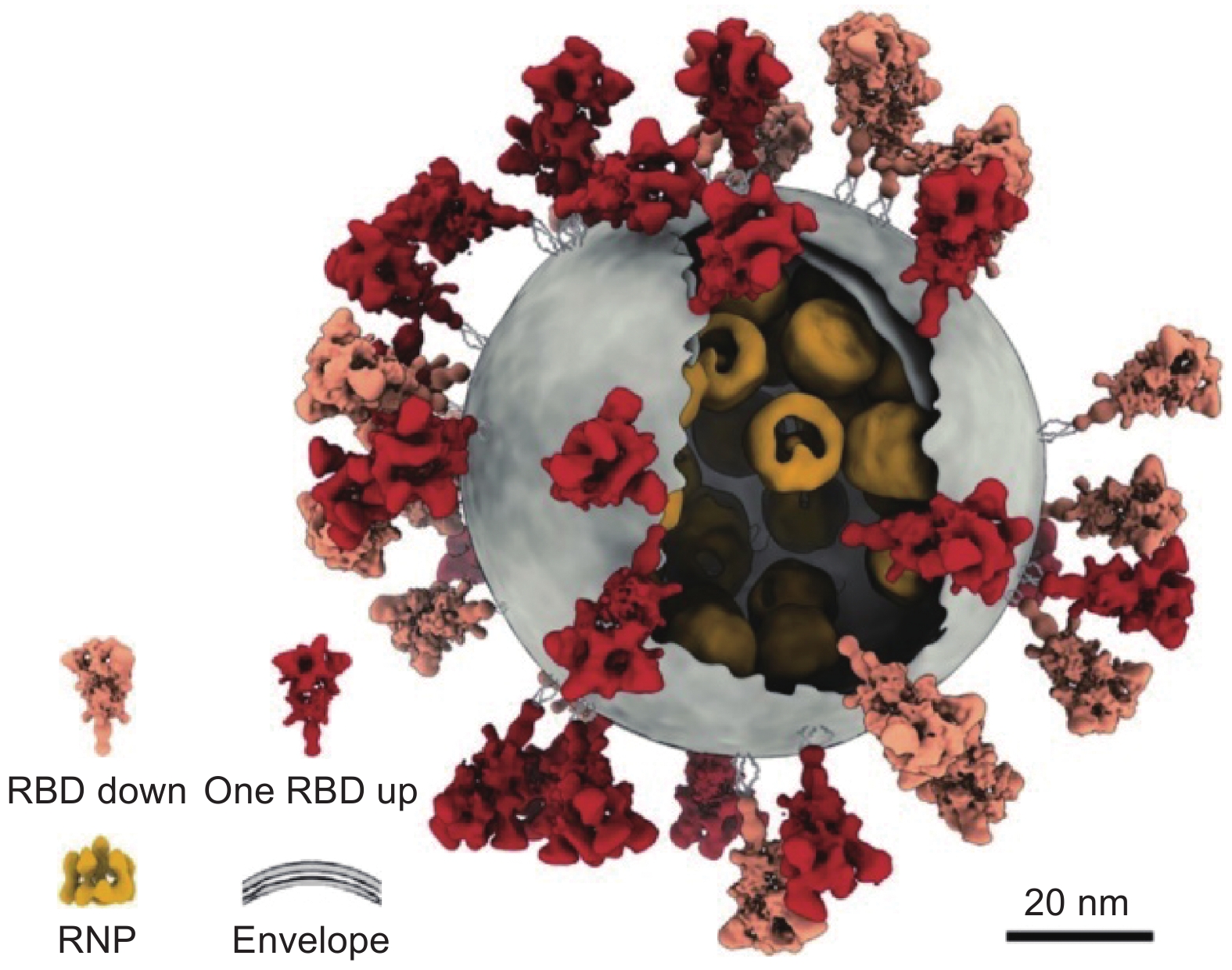
Yao H, Song Y, Chen Y, et al. Molecular Architecture of the SARS-CoV-2 Virus[J]. Cell,2020,183(3):730-738. doi: 10.1016/j.cell.2020.09.018
|
| [81] |
Bai R, Wan R, Yan C, et al. Mechanism of spliceosome remodeling by the ATPase/helicase Prp2 and its coactivator Spp2[J]. Science,2021,371(6525):eabe8863. doi: 10.1126/science.abe8863
|

Figures(14)
Address: 27 Taoyuan South Road, Yingze District, Taiyuan City, Shanxi Province, ChinaPostCode:030001
Phone/Fax:0351-2025254
Email: tcl@sxicc.ac.cn
Copyright © 2021 Editorial Office of New Carbon Materials. All Rights Reserved
This system consists of Beijing Renhe Information Technology Co. Ltd


 DownLoad: